Q.Draw the ray diagram of a compound microscope and show the tube length in it. (No description is required)
🔒You're viewing a preview — the full solution, concept, methods & PYQ mapping are locked.
🔒 Start your 14-day free trial to unlock the full solution →Concept understanding — Microscope Magnification
Microscope Magnification
A compound microscope views very small, nearby objects using two lenses in sequence: the objective (near the object) and the eyepiece (near the eye). Its total magnifying power is the product of what each lens contributes.
How the two lenses work together
- The object sits just beyond the focus of the short-focal-length objective (fo), which forms a real, inverted, magnified image inside the tube.
- That real image falls just inside the focus of the eyepiece (fe), which acts as a simple magnifier, producing a large virtual, magnified final image for the eye.
Because each stage magnifies, the effects multiply:
M=mo×me
The objective's magnification
mo=uovo≈foL
where L is the tube length (roughly the distance between the objective's second focal point and the eyepiece's first focal point). The object sits close to fo, so this approximation holds for a well-designed microscope.
The eyepiece's magnification
The eyepiece behaves as a simple magnifier:
- Final image at the near point (D=25 cm, largest magnification):
me=1+feD
- Final image at infinity (relaxed eye, "normal adjustment"):
me=feD
Total magnifying power
Image at the near point: M=foL(1+feD)
Image at infinity: M=foL⋅feD
High magnification needs short fo and fe (both sit in denominators) and a large tube length L — this is why a microscope objective is always a very short-focus lens.
Worked example
Objective fo=1.0 cm, eyepiece fe=2.5 cm, tube length L=20 cm, near point D=25 cm. Find M with the final image at the near point.
mo=1.020=20,me=1+2.525=11
M=20×11=220 …
Why this formula?
Microscope Magnification: Why the Formula Holds
Let's build the understanding from first principles — not just memorizing formulas, but seeing why they work.
1. What Does "Magnification" Mean in a Microscope?
A microscope creates a larger apparent image of a tiny object. The total magnification is the product of two stages:
- Objective lens — creates a real, enlarged, inverted image of the specimen.
- Eyepiece (ocular) — acts as a simple magnifier to view that real image.
So:
Total magnification = (magnification by objective) × (magnification by eyepiece)
2. The Key Formula
For a compound microscope in normal adjustment (final image at infinity, relaxed eye):
M=Mo×Me=(foL)×(feD)
Where:
- fo = focal length of objective
- fe = focal length of eyepiece
- L = tube length (distance between second focal point of objective and first focal point of eyepiece)
- D = near point distance of the eye (usually 25 cm)
3. Derivation of Objective Magnification Mo
Step 1: How the objective works
The objective lens forms a real, inverted, enlarged image of the specimen. The specimen is placed just outside its focal point (fo).
Step 2: Using the lens formula
For a thin lens:
vo1−uo1=fo1
(Using sign convention: uo is negative, vo is positive)
Step 3: The tube length approximation
In a standard microscope, the specimen is placed very close to fo, so:
- uo≈−fo (object just beyond focal point)
- The image is formed at the first focal point of the eyepiece, which is at a distance L from the second focal point of the objective.
Thus:
vo≈fo+L
Step 4: Magnification formula
Lateral magnification by objective:
Mo=∣uo∣vo≈fofo+L=1+foL
Since L≫fo in practice, 1 is negligible:
Mo≈foL
Why this makes sense: A shorter fo means the objective is more "powerful" — it bends light more sharply, creating a larger image at the fixed tube length.
4. Derivation of Eyepiece Magnification Me
Step 1: The eyepiece as a simple magnifier
The eyepiece takes the real image from the objective and acts like a magnifying glass. For relaxed eye (final image at infinity), the real image must be placed at the focal point of the eyepiece.
Step 2: Angular magnification
Angular magnification is defined as:
Me=angle subtended by object at near pointangle subtended by image at eye
For a simple magnifier with image at infinity:
Me=feD
Where D=25 cm (standard near point). …
A compound microscope has a short-focus objective forming a real, inverted, magnified image of a nearby object; the eyepiece then acts as a magnifier of that image. The tube length L is the distance between the back focal point of the objective and the front focal point of the eyepiece (the separation of the two lenses in the normal drawing). …
Object beyond F_o -> objective makes a real inverted magnified image -> eyepiece magnifies it to a final virtual image; the tube length L is the objective-to-eyepiece separation.
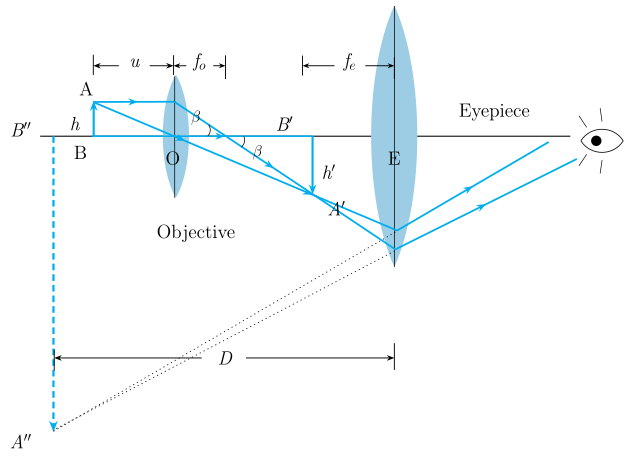
(A ray diagram is to be drawn; the description below states exactly what to draw and label.)
How to draw and read the compound microscope ray diagram:
- Place the small object AB just beyond the principal focus Fo of the objective lens (short focal length fo).
- Draw two rays from the tip B: one parallel to the axis (refracts through the far focus of the objective) and one through the optical centre of the objective (undeviated). They meet to form a real, inverted, magnified image A'B' inside the tube, lying just inside the focus of the eyepiece.
- This intermediate image A'B' now acts as the object for the eyepiece (short focal length fe), which is used as a simple magnifier. Draw rays from A'B' through the eyepiece; the refracted rays diverge, so trace them back to locate the final image, which is virtual, inverted (relative to AB) and highly magnified, formed at the least distance of distinct vision (or at infinity in normal adjustment). …
Showing the 12 most recent of 15 on this concept.
- CBSE 2026Set A1 markMCQQ.When the tube length of microscope is increased, its magnifying power (A) increases (B) decreases (C) becomes zero (D) remains unchanged
›Reveal solutionSolution
The magnifying power of a compound microscope is proportional to the tube length, so increasing L increases the magnification.
For a compound microscope the magnifying power is approximately
M=foL⋅feD …
- CBSE 2026Set ANNUAL1 markMCQQ.The image formed by a simple microscope is(a) imaginary and erect(b) imaginary and inverted(c) real and erect(d) real and inverted
›Reveal solutionSolution
A simple microscope is just a convex lens used with the object inside its focal length, which always produces a virtual, erect, magnified image.
A simple microscope is a single convex lens. When the object is placed BETWEEN the lens and its focal point (object distance less than f), the convex lens produces an image that is on the SAME side as the object, virtual (cannot be caught on a screen - here called 'imaginary'), erect (same orienta …
- CBSE 2026Set ANNUAL1 markMCQQ.Assertion (A): The magnifying power of a simple microscope is inversely proportional to the focal length of the lens. Reason (R): Power of a lens is inversely proportional to the focal length.(a) Both Assertion (A) and Reason (R) are true and Reason (R) is the correct explanation of Assertion (A).(b) Both Assertion (A) and Reason (R) are true, but Reason (R) is not the correct explanation of Assertion (A).(c) Assertion (A) is true, but Reason (R) is false.(d) Assertion (A) and Reason (R) both are false.
›Reveal solutionSolution
Both statements are true, and since M=D/f=D×P, the fact that power P∝1/f directly explains why magnifying power is inversely proportional to f.
For a simple microscope, magnifying power (image at infinity) is M=fD, where D is the least distance of distinct vision. This can be rewritten as M=D×f1=D×P, where P=f1 is the power of the lens. So M is directly proportional to the lens's power, and since power is inver …
- CBSE 2025Set X11 markMCQQ.Final image of a real object formed by a compound microscope is __________ with respect to the object.(a) real, inverted and magnified(b) virtual, erect and magnified(c) virtual, erect and diminished(d) virtual, inverted and magnified
›Reveal solutionSolution
(d) virtual, inverted and magnified. In a compound microscope the objective forms a real, inverted, magnified image, which acts as the object for the eyepiece. The eyepiece then forms a final imag …
- CBSE 2025Set IMPROVEMENT1 markQ.Write the formula for the total magnification of a compound microscope when the final image is formed at infinity.
›Reveal solutionSolution
For a compound microscope with the final image at infinity, the total magnifying power is the product of the objective's linear magnification and the eyepiece's angular magnification.
In a compound microscope, the objective lens forms a real, magnified image of the object; this image acts as the object for the eyepiece, which forms the final image. When the eyepiece is adjusted so that the final image is formed at infinity (normal/relaxed-eye viewing), the total magnifying power is:
M=mo×me=foL×feD …
- CBSE 2024Set A1 markMCQQ.Image formed in compound microscope is (A) real and erect (B) real and inverted (C) virtual and inverted (D) virtual and erect
›Reveal solutionSolution
A compound microscope forms a final image that is virtual, magnified and inverted → option (C).
In a compound microscope the objective lens first forms a real, inverted, magnified image of the object. This intermediate image acts as the object for the eyepiece, which is used as a simple magnifier and forms the **final image that is virtual, further magnifie …
- CBSE 2024Set ANNUAL1 markMCQQ.If the magnification of objective and eyepiece in a compound microscope is 'm_o' and 'm_e' respectively, then the total magnifying power (m) of the microscope will be -(a) m_o + m_e(b) m_o - m_e(c) m_o . m_e(d) m_o / m_e
›Reveal solutionSolution
In a compound microscope, the objective forms a magnified real image which the eyepiece further magnifies, so the two magnifications multiply.
In a compound microscope, the objective lens forms a real, magnified, inverted image of the object with magnification mo. This image acts as the object for the eyepiece, which further magnifies it (acting like a simple magnifier) with magnification me.
…
- CBSE 2023Set ANNUAL1 markMCQQ.The image formed by a simple microscope is(1) imaginary and erect(2) imaginary and inverted(3) real and erect(4) real and inverted
›Reveal solutionSolution
A simple microscope is a single converging lens used with the object inside its focal length, producing a virtual, erect, magnified image.
When the object is placed within the focal length of a convex lens, the emergent rays diverge, and appear to come from a point behind the lens on the same side as the object …
- CBSE 2022Set I1 markMCQQ.Magnifying power of simple microscope is (A) M = 1 - D/f (B) M = 1 + D/f (C) M = 1 - f/D (D) M = 1 + f/D
›Reveal solutionSolution
Simple microscope (image at near point): M=1+fD.
A simple microscope is a single convex lens of focal length f used as a magnifier. When the final image is formed at the least distance of distinct vision D (≈25 cm), the angular magnification is
M=1+fD.
…
- CBSE 2022Set HE2171 markMCQQ.The focal length of eye in compound microscope ________ the focal length of the objective.(i) is less than(ii) is more than(iii) is equal(iv) None of these
›Reveal solutionSolution
The eyepiece focal length of a compound microscope is greater than the objective's focal length.
In a compound microscope, the objective lens is placed very close to the small object and has a very small focal length fo, so that it forms a highly magnified real, inverted, enlarged image close to the eyepiece. The eyepiece then acts like a simple magnifier and further magnifies this image; it is given a comparatively larger focal length fe so it can view the intermediate image comfortably while still giving good magnification. …
- CBSE 2022Set HE2171 markQ.Match the following. Column A item: 'Magnifying power of Compound Microscope'. Choose its matching entry from Column B:(a) (1 - D/f)(b) Hertz(c) Electromagnetic induction(d) (-v0/u0)(1 + D/fe)(e) V.I.(f) Hershel(g) Instrument of measuring current(h) Instrument of measuring potential(i) Polarisation of light(j) Unit of energy
›Reveal solutionSolution
The magnifying power of a compound microscope is M=(−u0v0)(1+feD), matching option (d).
The overall magnification of a compound microscope is the product of the linear magnification produced by the objective, mo=v0/u0, and the angular magnification produced by the eyepiece used as a simple magnifier with the final image at the near point, me=1+D/fe (D = least distance of distinct vision). This gives t …
- CBSE 2021Set A1 markMCQQ.The image formed by simple microscope is (A) Virtual and erect (B) Virtual and inverted (C) Real and erect (D) Real and inverted
›Reveal solutionSolution
A simple microscope produces a virtual, erect, magnified image.
A simple microscope is a single convex lens used as a magnifying glass. The object is placed within the focal length (between the lens and its focus), so the lens forms an image that is virtual, erect, and enlarged, on the same side as the object (typically at the n …
🎓Unlock everything free for 14 days
- ✓Full step-by-step solutions
- ✓Concept-first explanations
- ✓Methods, shortcuts & mistakes
- ✓PYQ mapping + timed mock tests
Full access for 14 days. No credit card required.