Biology · Ch 1 — Sexual Reproduction in Flowering Plants
Stamen, Microsporangium and Pollen Grain
Stamen, Microsporangium and Pollen Grain
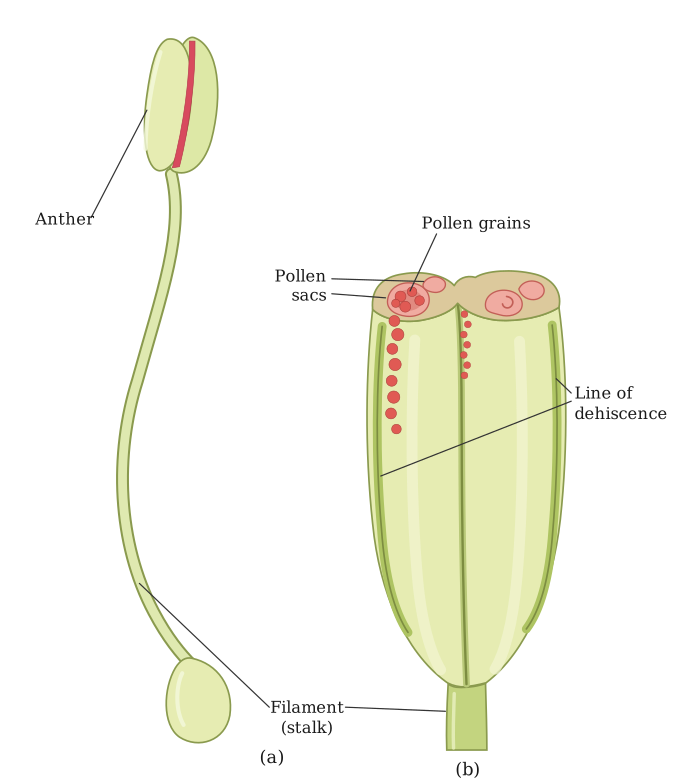
The stamen is the male reproductive unit of a flower. It consists of two parts: a long, slender stalk called the filament, and a terminal, generally bilobed structure called the anther. The filament is attached at its proximal end to the thalamus or to the petal of the flower.
The number and length of stamens vary greatly across different species. If you collect one stamen each from ten different flowers and arrange them on a slide, the large variation in size becomes obvious. Observing each stamen under a dissecting microscope reveals a range in the shape and attachment of anthers.
A typical angiosperm anther is bilobed, with each lobe having two theca — that is, it is dithecous. A longitudinal groove often runs lengthwise, separating the theca.
Structure of the Anther (Transverse Section)
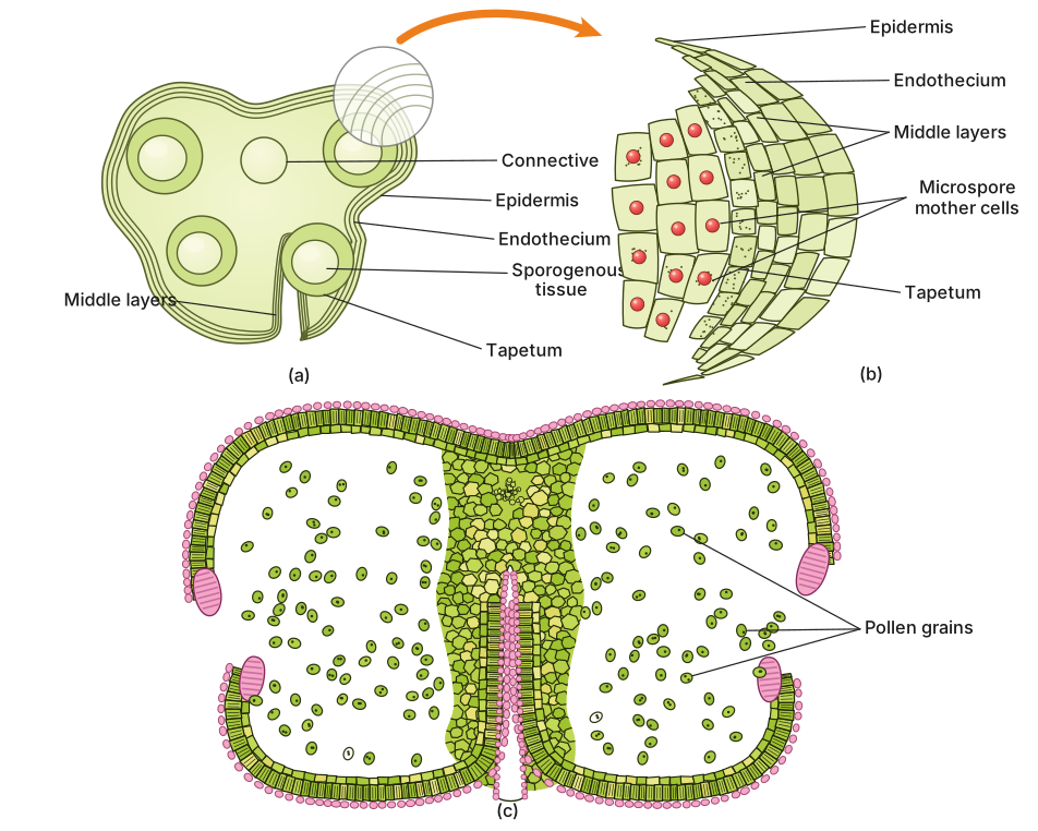
In transverse section, the bilobed nature of the anther is very distinct. The anther is a four-sided (tetragonal) structure containing four microsporangia located at the corners — two in each lobe. These microsporangia develop further and become pollen sacs. They extend longitudinally through the entire length of the anther and are packed with pollen grains.
Structure of a Microsporangium
In transverse section, a typical microsporangium appears nearly circular. It is surrounded by four wall layers:
- Epidermis — outermost layer.
- Endothecium — lies below the epidermis.
- Middle layers — one or more layers.
- Tapetum — the innermost layer.
The outer three wall layers (epidermis, endothecium, and middle layers) perform the functions of protection and help in the dehiscence (splitting open) of the anther to release pollen. The innermost layer, the tapetum, nourishes the developing pollen grains. Cells of the tapetum possess dense cytoplasm and generally have more than one nucleus (they are often binucleate).
(See the in-text question on tapetal cell nuclei, below.)
When the anther is young, the centre of each microsporangium is occupied by a group of compactly arranged, homogenous cells called the sporogenous tissue.
Microsporogenesis
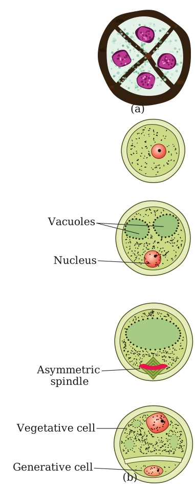
As the anther develops, the cells of the sporogenous tissue undergo meiotic divisions to form microspore tetrads. Each cell of the sporogenous tissue is capable of giving rise to a microspore tetrad, so each one is a potential pollen mother cell (PMC) or microspore mother cell.
The process of formation of microspores from a pollen mother cell through meiosis is called microsporogenesis. The microspores, as they are formed, are arranged in a cluster of four cells — the microspore tetrad. As the anthers mature and dehydrate, the microspores dissociate from each other and develop into pollen grains. Inside each microsporangium, several thousand microspores or pollen grains are formed, which are released when the anther dehisces.
(See the in-text question on tetrad ploidy, below.)
The Pollen Grain
Pollen grains represent the male gametophytes. They are generally spherical, measuring about 25–50 micrometers in diameter. The pollen grain has a prominent two-layered wall:
- Exine — the hard outer layer.
- Intine — the inner layer.
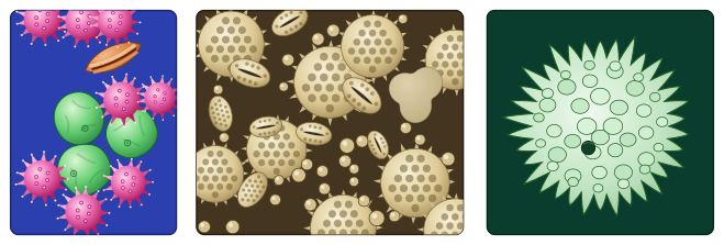
Exine: Made up of sporopollenin, one of the most resistant organic materials known. It can withstand high temperatures and strong acids and alkali. No enzyme that degrades sporopollenin is known so far. Because of sporopollenin, pollen grains are well-preserved as fossils. The exine exhibits a fascinating array of patterns and designs. It has prominent apertures called germ pores where sporopollenin is absent. (See the in-text question on why the exine is hard and what germ pores do, below.)
Intine: A thin and continuous layer made up of cellulose and pectin.
The cytoplasm of the pollen grain is surrounded by a plasma membrane. When the pollen grain is mature, it contains two cells:
- Vegetative cell — bigger, has abundant food reserve and a large, irregularly shaped nucleus.
- Generative cell — small, floats in the cytoplasm of the vegetative cell. It is spindle-shaped with dense cytoplasm and a nucleus.
In over 60 per cent of angiosperms, pollen grains are shed at this 2-celled stage. In the remaining species, the generative cell divides mitotically to give rise to two male gametes before pollen grains are shed — this is the 3-celled stage.
Pollen Viability and Storage
Pollen grains of many species cause severe allergies and bronchial afflictions in some people, often leading to chronic respiratory disorders like asthma and bronchitis. Parthenium (carrot grass), which came to India as a contaminant with imported wheat, has become ubiquitous and causes pollen allergy. …
Drawn by us to help you understand the concept clearly, and verified to make sure it's accurate. For exams, practice from your NCERT textbook's own diagram.
Figure 1.2 has two panels, (a) and (b), and together they give you the complete external and internal anatomy of the stamen — the male reproductive unit of a flower.
Panel (a) shows a single, complete stamen. You see the long, slender stalk called the filament, which attaches at its base to the thalamus or petal. At the tip of the filament sits the anther, which is clearly drawn as a bilobed structure — two distinct lobes side by side. This is the external view: a stalk topped by a two-lobed head. The caption calls it "a typical stamen," and the key takeaway is that the anther is not a single sac but a paired structure.
Panel (b) is a three-dimensional cut-away view of that anther. The artist has sliced through the anther to reveal its internal organisation. You can see that each of the two lobes is further divided into two chambers — these are the theca (singular: theca). Because each lobe has two theca, the anther is described as dithecous. Running lengthwise through each theca is a microsporangium (a pollen sac). Since there are two theca per lobe and two lobes, the anther contains four microsporangia in total — making it tetrasporangiate. Inside these four elongated chambers, the drawing shows numerous tiny dots or circles representing the pollen grains packed inside.
The figure also implies a relationship between the two panels: the external bilobed shape in (a) is the same structure whose interior is exposed in (b). A longitudinal groove that separates the theca on each lobe is often visible in such diagrams, though the description does not specify whether it is labelled. …
Drawn by us to help you understand the concept clearly, and verified to make sure it's accurate. For exams, practice from your NCERT textbook's own diagram.
Figure 1.3 is a three-panel diagram that walks you through the internal structure of an anther, from its young, undifferentiated state to the moment it splits open to release pollen. It is the visual companion to the text on microsporangium structure and dehiscence.
Panel (a) shows a transverse section (T.S.) of a young anther. The cut is taken across the anther while it is still developing, before any pollen has formed. The key feature here is the four microsporangia — one at each corner of the roughly tetragonal (four-sided) anther. Because the anther is bilobed, two microsporangia lie in each lobe. At this stage, the centre of each microsporangium is filled with sporogenous tissue, a mass of compact, homogenous cells that will later undergo meiosis. The panel also shows the bilobed outline of the anther, with a groove (the longitudinal groove) separating the two thecae of each lobe.
Panel (b) is an enlarged view of a single microsporangium from panel (a). It zooms in to reveal the four concentric wall layers that surround the central sporogenous tissue. From outermost to innermost, these are:
- Epidermis — the outermost protective layer.
- Endothecium — a layer that develops fibrous thickenings and helps in anther dehiscence.
- Middle layers — one to a few layers of cells that are crushed as the anther matures.
- Tapetum — the innermost layer, with dense, often binucleate cytoplasm. Its job is to nourish the developing pollen grains.
The centre of the microsporangium is occupied by the sporogenous tissue, which will give rise to microspore mother cells.
Panel (c) shows a mature, dehisced anther. The anther has now split open along the lines of dehiscence (typically at the junction of the two thecae in each lobe), exposing the pollen grains (the mature microspores) that fill each lobe's cavity. The figure captures the moment of release itself, with the grains still visible packed inside the burst-open sacs -- not an anther already emptied. This panel illustrates the final step in the sequence: the release of male gametophytes for pollination. …
Drawn by us to help you understand the concept clearly, and verified to make sure it's accurate. For exams, practice from your NCERT textbook's own diagram.
Figure 1.4 is a composite of several scanning electron micrographs (SEMs), each showing a single pollen grain from a different plant species. The caption tells you these are "a few pollen grains," and the surrounding text makes clear the purpose: to show the astonishing variety in size, shape, colour, and — most importantly — surface ornamentation that exists among pollen grains.
Each panel in the figure is a separate micrograph. There are no arrows, labels, or scale bars described in the textbook, so you should assume each image simply presents one pollen grain in isolation, at high magnification. The grains are not all the same shape: some are spherical, others are oval or elongated. The key teaching point is the exine — the hard outer wall made of sporopollenin. In these SEMs, the exine is not smooth; it is sculptured. You see ridges, spines, pits, grooves, and net-like patterns. This is the "fascinating array of patterns and designs" the text mentions. The germ pores — apertures where sporopollenin is absent — may appear as smooth, sunken spots or furrows on the surface of some grains, but the description does not specify which panels show them.
What this figure teaches you is that pollen grains are not just uniform yellow dust. Their external architecture is species-specific and remarkably intricate. The exine's sculpturing is so distinctive that it can be used to identify plant species, even from fossilised pollen. The figure directly supports the text's statement that the exine is hard and resistant — you are seeing a durable, preserved structure, not a delicate living cell. The variety also hints at different modes of pollination: spiny or sticky pollen is often insect-pollinated, while smooth, dry pollen is more typical of wind-pollinated plants, though the figure itself does not label which is which. …
Drawn by us to help you understand the concept clearly, and verified to make sure it's accurate. For exams, practice from your NCERT textbook's own diagram.
Figure 1.5 has two panels, (a) and (b), and together they show the entire journey from the products of meiosis to a mature male gametophyte.
Panel (a) is labelled "Enlarged view of a pollen grain tetrad". It shows a cluster of four haploid microspores still held together after meiosis of one pollen mother cell. This is the microspore tetrad — the immediate result of microsporogenesis. The four cells are identical in ploidy (haploid, n) and are enclosed within the callose wall that temporarily binds them. In the anther, thousands of such tetrads are formed inside each microsporangium. The panel makes clear that the tetrad is a transient stage: as the anther matures and dehydrates, these four microspores separate from each other.
Panel (b) is labelled "stages of a microspore maturing into a pollen grain". It shows a sequence — typically from left to right or top to bottom — of a single microspore developing into a mature pollen grain. The first stage is a freshly released microspore: a single cell with a thin wall and a central nucleus. The next stage shows the microspore enlarging and its wall differentiating into two distinct layers. The outer layer is the exine, a hard, resistant layer made of sporopollenin. The exine is not continuous — it has gaps called germ pores (apertures) where sporopollenin is absent. The inner layer is the intine, a thin, continuous layer of cellulose and pectin. The cytoplasm is bounded by a plasma membrane.
The final stage in the sequence shows the mature pollen grain. Inside, two cells are visible: a large vegetative cell (with abundant food reserves and a large, irregularly shaped nucleus) and a smaller generative cell (spindle-shaped, with dense cytoplasm and its own nucleus) that floats within the cytoplasm of the vegetative cell. This is the 2-celled stage at which pollen is shed in over 60% of angiosperms. In the remaining species, the generative cell would divide mitotically to produce two male gametes before shedding, giving a 3-celled pollen grain — but the figure as described shows the 2-celled stage.
The figure does not show the 3-celled stage. It stops at the 2-celled stage, which is the more common condition at the time of pollen release. …
Drawn by us to help you understand the concept clearly, and verified to make sure it's accurate. For exams, practice from your NCERT textbook's own diagram.
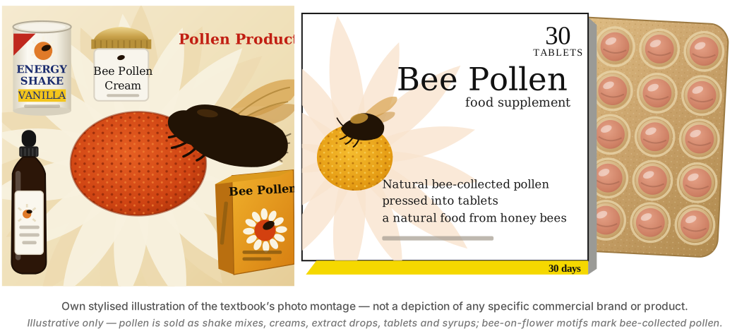
Figure 1.6, titled "Pollen products," is a small panel that sits alongside the boxed discussion on pollen as a nutritional supplement. It shows three separate items arranged side by side, each representing a commercial form in which pollen is sold for human consumption.
On the left, there is a jar or container labelled as pollen tablets — these are compressed, solid pellets made from dried pollen grains. In the centre, a bottle of pollen syrup is depicted, a liquid preparation where pollen is suspended or dissolved. On the right, a spoonful of loose pollen grains is shown, likely to illustrate the raw material itself. The overall layout is simple: no arrows or relationships are drawn between the items; they are just presented as a set of products derived from pollen.
The figure is not about the structure of a pollen grain or its development. Instead, it directly supports the textbook's point that pollen grains are "rich in nutrients" and are used as food supplements. The caption "Pollen products" makes this clear. The surrounding text also mentions that pollen can cause allergies and that its viability is short-lived in many species, but the figure itself does not show any of that — it only shows the finished consumer goods.
The figure is purely illustrative of the applied use of pollen, not of its biology. It is placed in the section on the stamen and pollen grain, but its purpose is to show a real-world application, not to teach anatomy or development. …