Chemistry · Ch 10 — Biomolecules
Structure of Proteins
Structure of Proteins
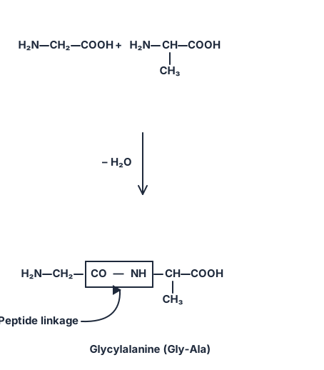
Proteins are polymers of α-amino acids, and the individual amino acid units are joined to each other through a peptide bond (also called a peptide linkage). Chemically, a peptide linkage is simply an amide formed between the group of one amino acid and the group of another.
When two amino acid molecules — either the same amino acid or two different ones — react, the amino group of one molecule combines with the carboxyl group of the other. This condensation eliminates a molecule of water and creates the peptide bond . The two-amino-acid product formed this way is called a dipeptide. For example, the carboxyl group of glycine reacting with the amino group of alanine gives the dipeptide glycylalanine:
Drawn by us to help you understand the concept clearly, and verified to make sure it's accurate. For exams, practice from your textbook's own diagram.
Redrawn from the NCERT page with the structures, printed labels (H2N, CH2, COOH, CH, CH3, – H2O, CO, NH, Peptide linkage) and reagent placement exactly as the textbook prints them. Every element of this display was checked against the printed page during the sweep's blind-judge verification pass, …
If a third amino acid attaches itself to a dipeptide, the product is a tripeptide, containing three amino acid units joined by two peptide linkages. In the same way, chains of four, five, or six amino acids are called tetrapeptides, pentapeptides, or hexapeptides respectively. Once more than ten amino acids are linked together, the product is termed a polypeptide. A polypeptide is called a protein once it has more than about a hundred amino acid residues and a molecular mass exceeding 10,000 u — though this distinction is not razor-sharp, and a shorter polypeptide chain is often still called a protein if it already has a well-defined protein-like conformation (insulin, for instance, is regarded as a protein even though it has only 51 amino acid residues).
Classification by Molecular Shape
Based on their overall molecular shape, proteins fall into two broad classes.
Fibrous proteins. When polypeptide chains run parallel to one another and are held together by hydrogen and disulphide bonds, the result is a thread-like, fibre-like structure. Fibrous proteins are generally insoluble in water. Keratin (found in hair, wool and silk) and myosin (found in muscles) are common examples.
Globular proteins. Here the polypeptide chains coil up around themselves to give a compact, roughly spherical shape. Globular proteins are usually soluble in water. Insulin and the various albumins are common examples.
Fibrous vs globular, at a glance
- Fibrous: parallel chains + H-bonds/disulphide bonds → thread-like → water-insoluble (keratin, myosin)
- Globular: chains coiled into a compact sphere → water-soluble (insulin, albumins)
The Four Levels of Protein Structure
The structure and overall shape of a protein can be studied at four progressively more complex levels — primary, secondary, tertiary and quaternary — with each level building on, and being more elaborate than, the one before it.
(i) Primary Structure
A protein may consist of one or more polypeptide chains. Within each polypeptide chain, the amino acids are linked to one another in a specific, defined sequence, and it is exactly this sequence that constitutes the primary structure of that protein.
The primary structure is extremely sensitive to change: altering even a single amino acid anywhere along this sequence produces, in effect, a completely different protein, since the identity and biological character of a protein are dictated by the precise order in which its amino acids are strung together.
(ii) Secondary Structure
The secondary structure describes the shape/conformation that a long polypeptide chain adopts. This shape arises from the regular folding of the polypeptide backbone, driven by hydrogen bonding between the (carbonyl) group and the (amide) group of the peptide bond. Polypeptide chains are found to exist in two principal types of secondary structure.
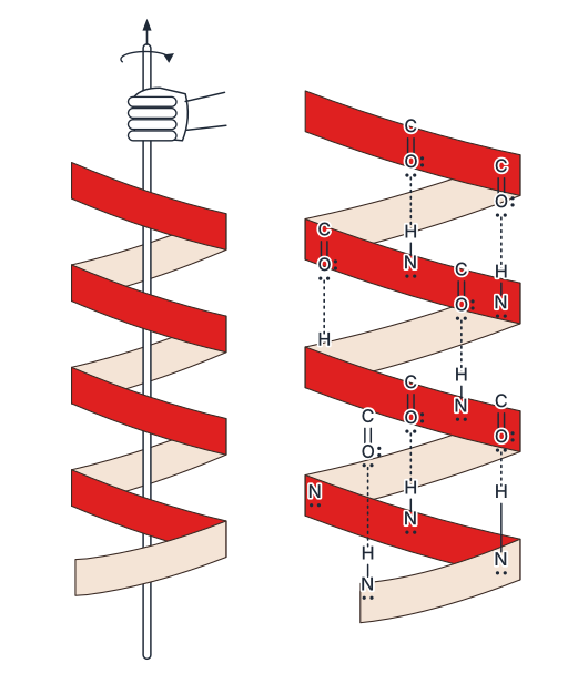
α-Helix. The α-helix is one of the most common ways in which a polypeptide chain arranges itself to form all the hydrogen bonds it possibly can. The chain twists into a right-handed screw (helix), with the group of every amino acid residue hydrogen-bonded to the group of an adjacent turn of the helix (see fig-10-1). In this arrangement:
- the polypeptide backbone itself forms the coiled helical spiral,
- the amino-acid side chains (R groups) project outward from the helix,
- and the structure is held together by intramolecular hydrogen bonds running along the length of the chain, linking each residue to the residue four positions further along.
Drawn by us to help you understand the concept clearly, and verified to make sure it's accurate. For exams, practice from your textbook's own diagram.
The α-helix is a common secondary structure of proteins. The figure depicts a single polypeptide chain coiled into a right-handed helix around an imaginary central axis. The backbone of the chain forms the spiral, while the R groups (side chains) of each amino acid project outward from the helix, away from the core.
The key structural feature shown is the pattern of intramolecular hydrogen bonds (typically drawn as dotted lines). Every backbone N–H group of one amino acid forms a hydrogen bond with the C=O group of the amino acid four residues ahead in the chain. This regular, repeating pattern — — locks the coil into a rigid, stable shape. The hydrogen bonds run parallel to the helix axis and are nearly straight, making them strong.
The figure teaches that the α-helix is stabilized entirely by these regularly spaced hydrogen bonds between peptide backbone atoms, not by interactions between side chains. The side chains point outward, so they are free to participate in higher-order folding (tertiary structure) or interactions with other molecules.
The textbook does not derive a specific formula from this figure, but the structural principle can be expressed in terms of the hydrogen bond pattern:
- Each turn of the helix contains approximately 3.6 amino acid residues.
- The rise per residue along the helix axis is about 1.5 Å.
- The pitch (distance for one complete turn) is therefore: …
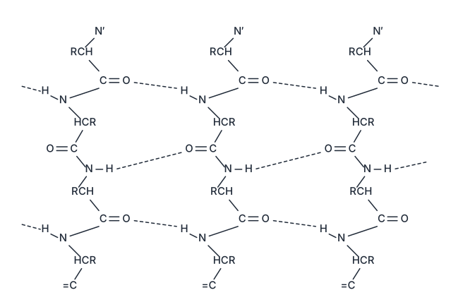
β-Pleated sheet. In the β-pleated sheet structure, the peptide chains are stretched out to nearly their maximum extension and then laid side by side, held together by intermolecular hydrogen bonds (i.e., hydrogen bonds between neighbouring chains rather than within a single chain, see fig-10-2). Because the resulting zig-zag arrangement of extended chains resembles the pleated folds of drapery or cloth, this structure is called the β-pleated sheet.
Drawn by us to help you understand the concept clearly, and verified to make sure it's accurate. For exams, practice from your textbook's own diagram.
The β-pleated sheet is the second major type of secondary structure in proteins. In this arrangement, the polypeptide backbone is almost fully extended — it is not coiled like the α-helix. Instead, two or more such extended chains lie side by side.
The figure shows these chains running in opposite directions (antiparallel). The backbone of each chain zig-zags, giving the sheet a pleated appearance, like the folds of a curtain or drapery. The side chains (R groups) of the amino acids project above and below the plane of the sheet.
The key structural feature is the hydrogen bonds that stitch the neighbouring chains together. These bonds form between the N–H group of one chain and the C=O group of the adjacent chain. In the diagram, these bonds are shown as dotted lines connecting the backbone atoms of different chains.
Physical idea: The β-pleated sheet is stabilised entirely by intermolecular hydrogen bonds between the peptide backbones of separate polypeptide strands (or between distant segments of the same chain that fold back). This is different from the α-helix, where hydrogen bonds are intramolecular (within the same chain, between turns of the helix). …
α-Helix vs β-pleated sheet
- α-Helix: a single chain coils into a right-handed spiral; backbone forms the helix, R groups point outward; stabilised by intramolecular H-bonds between every 4th residue.
- β-Pleated sheet: extended, zig-zag chains lie side by side; stabilised by intermolecular H-bonds between adjacent chains, giving a flat, sheet-like arrangement.
(iii) Tertiary Structure
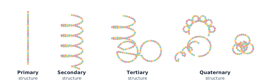
The tertiary structure represents the overall three-dimensional folding of the entire polypeptide chain — that is, a further level of folding superimposed on the secondary structure, describing how the helical and/or pleated-sheet segments of a chain pack together in space to give the molecule its overall shape (see fig-10-3, which diagrams all four levels together with each coloured ball representing an amino acid).
Drawn by us to help you understand the concept clearly, and verified to make sure it's accurate. For exams, practice from your textbook's own diagram.
Figure 10.3 is a schematic diagram that illustrates the four levels of protein structure using a simplified bead-and-string model. Each coloured bead represents a single amino acid, and the string connecting them is the peptide bond backbone.
- Primary structure is shown as a straight, linear sequence of beads. This represents the specific order of amino acids in the polypeptide chain. No folding or coiling is present — it is the simplest level.
- Secondary structure is depicted as the same chain coiled into a regular helix (specifically the -helix). This coiling arises from hydrogen bonds between the and groups of the peptide backbone, stabilising the helical shape.
- Tertiary structure shows the entire polypeptide chain folded and compacted into a three-dimensional globular shape. This folding results from interactions between side chains: hydrogen bonds, disulphide linkages (), van der Waals forces, and electrostatic attractions.
- Quaternary structure presents two or more folded subunits (each a separate tertiary structure) associating together. The figure specifically shows two subunits of two different types (e.g., the and chains of haemoglobin), held together by the same non-covalent forces.
Physical idea taught: The figure emphasises that protein structure is hierarchical — each level builds upon the previous one. The primary sequence determines how the chain can fold into secondary structures, which then pack into a tertiary shape, and multiple such shapes may assemble into a quaternary complex. Any change in the primary sequence (e.g., a single amino acid substitution) can alter all higher levels and destroy biological activity.
Key formula(s) developed with this figure: The textbook does not introduce a single formula here, but the concept of denaturation is linked to the disruption of stabilising interactions. The energy required to break a hydrogen bond is approximately:
…
It is the tertiary structure that gives rise to the two major molecular shapes discussed above — fibrous and globular. The principal forces that stabilise both the secondary and tertiary structures of a protein are:
- hydrogen bonds,
- disulphide linkages,
- van der Waals forces, and
- electrostatic (ionic) forces of attraction.
(iv) Quaternary Structure …
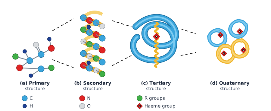
Drawn by us to help you understand the concept clearly, and verified to make sure it's accurate. For exams, practice from your textbook's own diagram.
What Figure 10.4 Shows
The figure is a four-panel diagram (a)–(d) that builds the structure of haemoglobin from the simplest to the most complex level. Each panel corresponds to one of the four structural levels of proteins described in the text.
-
Panel (a) – Primary structure: A single amino-acid unit is shown as a ball-and-stick model. This represents the fundamental building block of the polypeptide chain. The primary structure is the specific sequence of amino acids linked by peptide bonds. Any change in this sequence creates a different protein.
-
Panel (b) – Secondary structure: A helical stretch is depicted with a ribbon backbone. This is the -helix, one of the two common secondary structures. It arises from regular folding of the polypeptide backbone due to hydrogen bonding between the group of one peptide bond and the group of another, four residues away. The other secondary structure, the -pleated sheet, is not shown here but is described separately.
-
Panel (c) – Tertiary structure: One folded sub-unit (a single polypeptide chain) is shown as a ribbon, with a red haem group (the oxygen-binding site) held within. This represents the overall three-dimensional folding of the secondary structure into a globular shape. The forces stabilising this level include hydrogen bonds, disulphide linkages (), van der Waals forces, and electrostatic attractions.
-
Panel (d) – Quaternary structure: Four sub-units (two of each type, typically two and two chains) are assembled together, each carrying a haem group. This is the spatial arrangement of multiple polypeptide chains (sub-units) with respect to each other. Haemoglobin is a classic example of a protein with quaternary structure.
Arrows connect the panels sequentially, showing that each level is built upon the previous one.
Physical Idea Taught
The figure teaches that protein structure is hierarchical. The function of a protein (e.g., oxygen transport by haemoglobin) depends on its precise three-dimensional shape, which emerges from the sequence of amino acids (primary) through local folding (secondary) to global folding (tertiary) and finally assembly of multiple chains (quaternary). Disruption of any level — especially the hydrogen bonds that stabilise secondary and tertiary structures — leads to denaturation, where the protein loses its biological activity.
Key Formula(s) from the Textbook
The textbook does not develop a single formula directly from this figure. Instead, it describes the forces that stabilise the higher-order structures. The key relationships are:
- Hydrogen bond energy (typical for -helix):
Each hydrogen bond between and groups contributes roughly this stabilisation.
- Disulphide linkage (covalent bond between cysteine residues):
This is a strong covalent bond (bond energy ) that locks tertiary structure.
- van der Waals interactions (between nonpolar side chains): …