Biology · Ch 7 — Human Health and Disease
Acquired Immunity
Acquired Immunity
Acquired immunity is fundamentally different from innate immunity. While innate immunity is a general, non-specific defence that works against any invader, acquired immunity is pathogen specific — it targets one particular pathogen and remembers it for the future.
The key feature that sets acquired immunity apart is memory. When your body meets a pathogen for the first time, it mounts a primary response. This first response is slow and of low intensity because the immune system has never seen this enemy before and needs time to figure out how to fight it.
But here is the crucial part: the body remembers that first encounter. If the same pathogen attacks again, the immune system launches a secondary response (also called an anamnestic response). This second response is highly intensified — much faster and stronger than the first. This memory is why many diseases, like chickenpox, usually only happen once in a lifetime.
The primary response is weak and slow; the secondary (anamnestic) response is strong and fast — this is the hallmark of immunological memory.
The Cells That Carry Out Acquired Immunity
The primary and secondary immune responses are carried out by two special types of lymphocytes (a kind of white blood cell) present in our blood:
- B-lymphocytes (B-cells): These cells produce an army of proteins called antibodies in response to pathogens. Antibodies are released into the blood to fight the invaders.
- T-lymphocytes (T-cells): These cells do not secrete antibodies themselves. Instead, they help B-cells to produce antibodies.
Structure of an Antibody
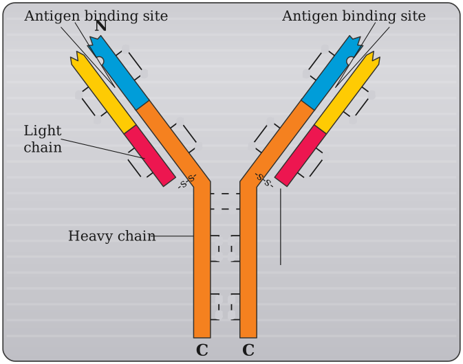
Each antibody molecule is made of four peptide chains:
- Two small chains called light chains
- Two longer chains called heavy chains
Because of this structure, an antibody is represented as H₂L₂ (two heavy chains and two light chains). Different types of antibodies are produced in our body, including IgA, IgM, IgE, and IgG.
Two Types of Acquired Immune Response
The acquired immune response is divided into two types:
- Humoral Immune Response (Antibody-mediated): Because antibodies are found in the blood (the "humors" of the body), this response is called humoral. It is mediated by B-cells and the antibodies they produce.
- Cell-mediated Immune Response (Cell-mediated immunity or CMI): This response is mediated by T-lymphocytes. It does not involve antibodies directly.
Why Organ Transplants Are Difficult: The Role of CMI
The cell-mediated immune response is responsible for a very practical problem in medicine: graft rejection. …
Drawn by us to help you understand the concept clearly, and verified to make sure it's accurate. For exams, practice from your textbook's own diagram.
The figure is a simplified cartoon of a single antibody molecule, drawn in the classic Y-shape that you will see in almost every textbook. The purpose is to show the basic quaternary structure — how the four protein chains are arranged and which parts do the work.
At the centre of the Y is a disulphide bond (a strong covalent link between sulphur atoms) that holds the two arms together. The molecule is built from four polypeptide chains: two identical heavy chains (longer, shown in one colour) and two identical light chains (shorter, shown in a different colour). Because there are two heavy and two light chains, the antibody is written as H₂L₂.
The figure also explicitly prints the N (amino) terminus near the tip of one arm and C (carboxyl) terminus labels at the base of each heavy-chain stem, alongside the Antigen binding site / Light chain / Heavy chain labels.
Each heavy chain runs from the tip of one arm, through the hinge region, and down into the stem of the Y. Each light chain sits alongside the upper part of a heavy chain, on the outside of the arm. The two arms are identical — the molecule is symmetric.
The most important feature is at the tips of the two arms. Here, the variable regions of one heavy chain and one light chain come together to form the antigen-binding site. This is the pocket that recognises and grabs onto a specific pathogen (antigen). Because there are two arms, each antibody can bind two antigens at once — this is called bivalency.
The stem of the Y (the constant region of the heavy chains) is the part that interacts with other immune cells and molecules. It determines which class the antibody belongs to (IgG, IgA, IgM, IgE) and triggers the rest of the immune response after the antigen is bound.
The figure does not show the hinge region as a separate label, but the flexible hinge between the arms and the stem is what allows the two antigen-binding sites to move and grab antigens that are spaced apart on a pathogen’s surface. …