Question of 16
Q.Describe the structure of cardiac muscle.
Telangana TsbieTelangana Board of Intermediate Education (Intermediate 1st Year) 2019Subjective· 4mImportance★★★★★
0% · 0/16 Questions
You're viewing a preview — the full solution, concept, methods & PYQ mapping are locked.
Start your 14-day free trial to unlock the full solution →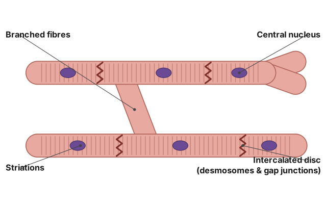
Cardiac muscle is a unique striated, involuntary muscle type restricted to the heart, whose branching fibres are electrically and mechanically joined by intercalated discs to enable coordinated, tireless, rhythmic contraction.
Location: Cardiac muscle forms the myocardium, the muscular middle layer of the heart wall, and is found nowhere else in the body.
Structure:
- Fibres are cylindrical and branched, forming an interconnected network.
- They show cross-striations (alternating light and dark bands), similar to skeletal muscle, due to the regular arrangement of actin and myosin filaments in sarcomeres.
- Each fibre is typically uninucleate (a single, centrally placed nucleus), occasionally binucleate.
- Fibres are joined end-to-end at specialised junctions called intercalated discs, which contain:
- Desmosomes — provide strong mechanical adhesion between adjacent cells.
- Gap junctions — allow direct electrical continuity, so action potentials spread rapidly from cell to cell.
- Numerous mitochondria are packed between myofibrils, reflecting the high, continuous energy demand of the heart.
Functional significance: …
Unlock everything free for 14 days
- Full step-by-step solutions
- Concept-first explanations
- Methods, shortcuts & mistakes
- PYQ mapping + timed mock tests
Full access for 14 days. No credit card required.