Muscular Tissue Types – From Intuition to Precision
Think about the last time you lifted a book, blinked, or felt your heart pound after a sprint. Each of those actions — voluntary, involuntary, and rhythmic — is powered by a different kind of muscle. The body doesn't use one universal muscle; it has three specialised versions, each built for a specific job.
The core idea is simple: structure determines function. Where a muscle is located, whether you can control it consciously, and how its cells are arranged all follow from what that muscle is meant to do.
The Three Types at a Glance
| Feature | Skeletal Muscle | Smooth Muscle | Cardiac Muscle |
|---|
| Location | Attached to bones | Walls of hollow organs (stomach, blood vessels, bladder, uterus) | Wall of the heart (myocardium) |
| Control | Voluntary (you decide to move) | Involuntary (autonomic nervous system + hormones) | Involuntary (autonomic + intrinsic pacemaker) |
| Cell shape | Long, cylindrical, unbranched | Spindle-shaped (tapered ends), no striations | Short, branched, cylindrical |
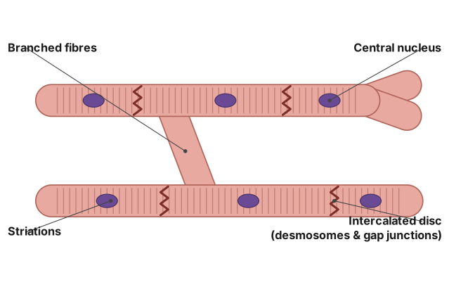
| Striations | Present (alternating light/dark bands) | Absent | Present |
| Nuclei | Multiple, peripheral | Single, central | Usually one or two, central |
| Intercalated discs | Absent | Absent | Present (unique to cardiac muscle) |
| Speed of contraction | Fast | Slow | Moderate, rhythmic |
| Fatigue | Fatigues easily | Does not fatigue | Does not fatigue (beats a lifetime) |
Skeletal Muscle – The Voluntary Worker
Skeletal muscle is what you think of when you picture "muscle" — biceps, quadriceps, the muscles that move your face. Its cells are long, unbranched cylinders packed with parallel myofibrils, which give it the striped (striated) appearance under a microscope. Each cell has many nuclei pushed to the edge, because the cell is so large.
The striations come from the precise arrangement of actin and myosin filaments into repeating units called sarcomeres. This organisation allows rapid, powerful contraction.
You control skeletal muscle consciously through the somatic nervous system. It contracts fast, generates force, but tires quickly — which is why you can't hold a heavy weight forever.
Smooth Muscle – The Unseen Regulator
Smooth muscle lines the walls of your stomach, intestines, blood vessels, airways, and bladder. Its cells are spindle-shaped — fat in the middle, tapering at both ends — with a single central nucleus. There are no striations because the actin and myosin filaments are not arranged in neat sarcomeres; they are scattered in a more irregular pattern.
A common mistake is to think "smooth" means it contracts smoothly. It does, but the name actually refers to the absence of striations under the microscope.
Smooth muscle is involuntary. You don't decide when your stomach churns or your blood vessels constrict. It contracts slowly and can stay contracted for long periods without tiring — essential for functions like digestion and blood pressure regulation.
Cardiac Muscle – The Tireless Pump
Cardiac muscle is found only in the heart. Its cells are short, branched, and cylindrical, with one or two central nuclei. Like skeletal muscle, it is striated — the sarcomeres are present. But unlike skeletal muscle, it is involuntary. …