Botany · Ch 11 — Microbes in Human Welfare
Microbes in Household Products
Microbes in Household Products
Curd, from milk
Curd is the simplest example of microbial transformation. Lactobacillus and other bacteria commonly grouped together as lactic acid bacteria (LAB) grow in milk and convert it to curd. As they grow, the LAB produce acids that coagulate and partially digest the milk proteins. In practice, a small amount of already-formed curd is added to fresh milk as a starter — it carries millions of LAB which, at a suitable temperature, multiply rapidly and convert the whole batch of milk to curd.
Curd made this way isn't just a change in texture: the fermentation also improves the milk's nutritional quality, increasing its vitamin B12 content. The same LAB continue to do useful work once eaten — inside our own stomach, they help keep disease-causing microbes in check.
Dough for idli, dosa, and bread
The dough used to make foods like dosa and idli is also fermented by bacteria. The puffed-up, aerated texture of the batter comes from the carbon dioxide gas the fermentation produces.
Bread depends on a different microbe entirely — the yeast Saccharomyces cerevisiae (baker's yeast), used to leaven the dough (see below).
Traditional fermented drinks and foods
A number of traditional Indian drinks and foods are made by fermentation. Toddy, a traditional drink in parts of southern India, is made by fermenting the sap collected from palms. Microbes are also used to ferment fish, soyabean, and bamboo shoots into foods.
Cheese
Cheese is one of the oldest food items made using microbes. The different varieties of cheese are known by their characteristic texture, flavour and taste — and that specificity comes from the particular microbes used to make each one.
- Swiss cheese gets its large holes from a bacterium called Propionibacterium sharmanii, which produces a large amount of carbon dioxide as it grows; the gas gets trapped in the cheese, forming the characteristic bubbles.
- Roquefort cheese is ripened by growing a specific fungus on it, which gives the cheese its particular flavour.
Swiss cheese's holes come from a bacterium; Roquefort's flavour comes from a fungus grown on the cheese. Both are examples of how the specific microbe used determines the character of the final product.
Bread: the leavening action of yeast
Bread depends entirely on a microbe — the yeast Saccharomyces cerevisiae, often called baker's yeast. When yeast is mixed into dough (flour and water), it ferments the sugars present in the flour. The main products of this fermentation are carbon dioxide gas and ethanol. …
Drawn by us to help you understand the concept clearly, and verified to make sure it's accurate. For exams, practice from your textbook's own diagram.
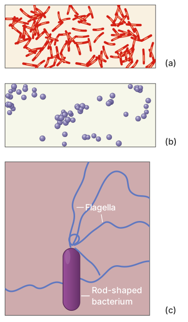
This figure is a set of three separate light-micrograph panels, each showing a different view of bacteria. The panels are labelled (a), (b), and (c), and they are arranged to help you compare bacterial shapes and structures.
Panel (a) shows rod-shaped bacteria, called bacilli, at a magnification of 1500X. You see elongated, cylindrical cells — like tiny pills or short rods — scattered across the field. This rod shape (bacillus) is common to many different bacterial genera -- the real caption doesn't identify which species this particular micrograph shows.
Panel (b) shows spherical bacteria, called cocci, also at 1500X. Here the cells appear as small, round dots, often clustered together. This spherical shape (coccus) is likewise common to many different bacterial genera -- again, the real caption doesn't name which species is pictured here.
Panel (c) is a much higher magnification — 50,000X — and focuses on a single rod-shaped bacterium. The key detail here is that you can see thin, thread-like structures called flagella extending from the surface of the cell. Flagella are the bacterial "tails" that allow movement (motility). At 1500X, flagella are too thin to be visible, so this panel uses a much higher magnification to reveal them.
The magnification numbers are important: 1500X means the image is 1500 times larger than the actual cell, while 50,000X is an electron-microscope-level magnification. This is why flagella are only visible in panel (c). …
Drawn by us to help you understand the concept clearly, and verified to make sure it's accurate. For exams, practice from your textbook's own diagram.
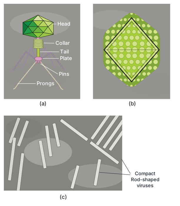
Figure 8.2 is a composite of three electron micrographs, each showing a different virus at a magnification of roughly 1,00,000–1,50,000 times their actual size. The three panels are labelled (a), (b), and (c), and they are arranged to compare the structural diversity of viruses.
Panel (a) shows a bacteriophage — a virus that infects bacteria. The image clearly reveals its distinctive structure: a polyhedral (many-sided) head that contains the genetic material, a rigid tail sheath attached to the head, and fine tail fibres extending from the base of the sheath. These fibres are what the phage uses to latch onto a bacterial cell before injecting its DNA.
Panel (b) displays an adenovirus, which causes respiratory infections in humans. Unlike the complex, tailed bacteriophage, the adenovirus particle appears as a roughly spherical, icosahedral (20-faced) structure. Its surface is made of protein subunits arranged in a symmetrical shell, and no tail or fibres are visible at this magnification — it is a much simpler, naked icosahedral virus.
Panel (c) shows the Tobacco Mosaic Virus (TMV), which infects plants. Its shape is completely different from the other two: it is an elongated, rigid rod. The rod is not smooth but appears to have a helical pattern, reflecting the way its protein subunits stack around the central RNA strand. TMV is the classic example of a helical virus.
The three panels together illustrate that viruses come in at least three fundamental shapes: complex (bacteriophage with head and tail), icosahedral (adenovirus), and helical/rod-shaped (TMV). This structural variety is directly linked to how each virus infects its host — the bacteriophage needs a tail to inject DNA through a bacterial wall, while adenovirus and TMV rely on different entry mechanisms suited to animal and plant cells, respectively. …
Drawn by us to help you understand the concept clearly, and verified to make sure it's accurate. For exams, practice from your textbook's own diagram.
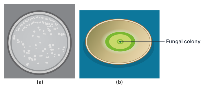
Figure 8.3 shows two separate petri dishes side by side, labelled (a) and (b). Both dishes contain a solid nutrient medium — typically agar — that supports microbial growth. In panel (a), you see several small, round, smooth-edged colonies scattered across the agar surface. These are bacterial colonies. Each colony started from a single bacterial cell that multiplied into a visible clump. The colonies are discrete and well-separated because bacteria tend to grow as compact, circular masses on solid media.
Panel (b) shows a very different appearance. Here, a single fuzzy, filamentous colony spreads irregularly across the plate. This is a fungal colony. The fuzzy texture comes from the network of thread-like hyphae that make up the fungal body. Unlike bacterial colonies, fungal colonies often grow outward in a radial pattern, covering a larger area and sometimes merging into a continuous mat. …