Botany · Ch 6 — Principles of Inheritance and Variation
Chromosomal Disorders
Chromosomal Disorders
Chromosomal Disorders: When Chromosomes Go Wrong
Mendelian disorders arise from mutations in single genes. Chromosomal disorders are fundamentally different — they are caused by the absence, excess, or abnormal arrangement of one or more whole chromosomes. Because entire chromosomes carry hundreds of genes, these disorders have widespread, often severe, effects on the individual.
How Do These Abnormalities Happen?
The textbook describes two main mechanisms:
-
Aneuploidy — This is the gain or loss of a single chromosome. It happens when chromatids fail to separate (non-disjunction) during cell division. The result is a cell with either one extra chromosome (trisomy) or one missing chromosome (monosomy).
-
Polyploidy — This is an increase in a whole set of chromosomes. It occurs when cytokinesis fails after telophase. Polyploidy is common in plants but rarely seen in animals.
A normal human cell has 46 chromosomes — 22 pairs of autosomes and one pair of sex chromosomes (XX in females, XY in males).
Trisomy and Monosomy
When an individual has an extra copy of any one chromosome, it is called trisomy (2n+1). When an individual lacks one chromosome from a pair, it is called monosomy (2n-1). Both situations lead to very serious consequences.
The Three Major Human Chromosomal Disorders
The NCERT textbook discusses three classic examples in detail.
1. Down's Syndrome (Trisomy 21)
- Cause: Presence of an additional copy of chromosome number 21 (trisomy of 21).
- First described by: Langdon Down in 1866.
- Key features:
- Short stature
- Small, round head
- Flat back of the head
- Broad, flat face
- Furrowed tongue and partially open mouth
- Broad palm with a characteristic single palm crease
- Many "loops" on fingertips
- Congenital heart disease
- Retarded physical, psychomotor, and mental development
2. Klinefelter's Syndrome
- Cause: Presence of an additional copy of the X chromosome, resulting in a karyotype of 47, XXY.
- Key features:
- Overall masculine development
- However, feminine development is also expressed — specifically, development of breasts (gynaecomastia)
- Tall stature with feminised character
- Individuals are sterile
3. Turner's Syndrome …
Drawn by us to help you understand the concept clearly, and verified to make sure it's accurate. For exams, practice from your textbook's own diagram.
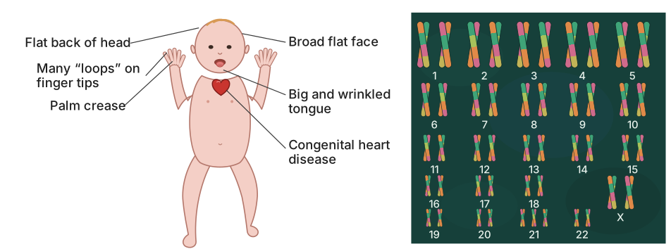
Figure 4.16 is a two-part illustration. The left side shows a full-body drawing of a person with Down’s syndrome, and the right side shows the corresponding karyotype (the complete set of chromosomes) of that individual.
The person is depicted with the characteristic physical features described in the textbook: short stature, a small round head, a broad flat face, a furrowed tongue, and a partially open mouth. The palm is drawn broad with a single deep crease across it (the simian crease). A label points to the heart, indicating congenital heart disease. The fingers are shown with many loops on the fingertips — another typical feature. The overall appearance is meant to represent the consistent set of traits that result from the underlying chromosomal abnormality.
The karyotype on the right is a standard arrangement of human chromosomes, numbered from 1 to 22 (the autosomes) plus the sex chromosomes. The key detail is that chromosome 21 appears three times instead of the normal two — this is trisomy 21. The total chromosome count is therefore 47, not 46. The sex chromosomes are shown as XX, indicating a female individual (though Down’s syndrome occurs in both sexes; the figure uses a female karyotype as a representative example). …
Drawn by us to help you understand the concept clearly, and verified to make sure it's accurate. For exams, practice from your textbook's own diagram.
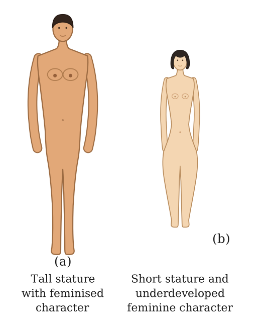
Figure 4.17 is a diagrammatic representation that places two human figures side by side for direct comparison. On the left, panel (a) shows a person with Klinefelter syndrome; on the right, panel (b) shows a person with Turner syndrome. The caption and the surrounding text make clear that both are genetic disorders caused by an abnormal number of sex chromosomes — not autosomes.
The Klinefelter figure (a) is drawn as a tall individual with masculine body build, but with one distinctly feminine feature: breast development (gynaecomastia). The text tells us the karyotype is 47, XXY — an extra X chromosome in a male. The figure conveys that despite the overall masculine development, the extra X leads to partial feminisation and sterility. No internal organs are shown; the figure simply illustrates the external, visible traits: tall stature and the presence of breasts.
The Turner figure (b) is drawn as a short-statured female. The text specifies the karyotype as 45, X0 — one X chromosome missing. The figure shows underdeveloped feminine secondary sexual characters (for example, lack of breast development and absence of typical female body contours). The text adds that the ovaries are rudimentary, so the individual is sterile. Again, only external features are depicted: short height and a lack of normal female secondary sexual traits.
The two panels are arranged so that the contrast is immediate: tall with feminised features versus short with underdeveloped feminine features. No arrows, labels for body parts, or additional annotations are described in the figure — just the two full-body silhouettes with their characteristic appearances. The figure’s purpose is to give a visual anchor for the two most common sex-chromosome aneuploidies in humans, making it clear that an extra X in a male produces a different set of physical outcomes than a missing X in a female. …