Q.Write the structures of fragments produced on complete hydrolysis of DNA. How are they linked in DNA molecule? Draw a diagram to show pairing of nucleotide bases in double helix of DNA.
🔒You're viewing a preview — the full solution, concept, methods & PYQ mapping are locked.
🔒 Start your 14-day free trial to unlock the full solution →Concept understanding — Nucleic Acid Components
Nucleic Acid Components: The Alphabet of Life
Imagine you want to write a book that contains all the instructions for building and running a living organism. You'd need an alphabet — a set of letters that can be combined in endless ways to form words, sentences, and chapters. In living cells, that alphabet is made of nucleic acids (DNA and RNA), and each "letter" is called a nucleotide.
The Big Picture: What Are Nucleic Acids?
Nucleic acids are long, chain-like molecules that store and transmit genetic information. DNA holds the master blueprint; RNA helps execute it. But both are built from the same basic building block: the nucleotide.
Think of a nucleotide as a single Lego brick. DNA and RNA are long chains of these bricks, each brick slightly different from the next.
The Three Parts of a Nucleotide
Every nucleotide has three components, like a three-part key:
- A phosphate group — a small, negatively charged group (PO43−). It acts like the "handle" that links nucleotides together.
- A sugar — either ribose (in RNA) or deoxyribose (in DNA). This is the "body" of the brick.
- A nitrogenous base — a ring-shaped molecule containing nitrogen. This is the "colored part" that carries the actual information.
The sugar and base together form a nucleoside. When you add the phosphate, you get a nucleotide.
Nucleoside = Sugar + Base
Nucleotide = Sugar + Base + Phosphate
The Two Families of Bases
The bases come in two structural types:
- Purines (double-ring structures): Adenine (A) and Guanine (G)
- Pyrimidines (single-ring structures): Cytosine (C), Thymine (T) (only in DNA), and Uracil (U) (only in RNA)
A mnemonic: Purines are Pure as All Gold (A and G). Pyrimidines are CUT (C, U, T).
DNA vs. RNA: The Key Differences
| Feature | DNA | RNA |
|---|---|---|
| Sugar | Deoxyribose (missing one oxygen) | Ribose (has that oxygen) |
| Bases | A, G, C, T | A, G, C, U |
| Structure | Double-stranded helix | Usually single-stranded |
| Function | Stores genetic information | Carries and executes instructions |
A common mistake: thinking "nucleoside" and "nucleotide" are the same. Remember: nucleotide has the phosphate; nucleoside does not. ATP (adenosine triphosphate) is a nucleotide — it's the energy currency of the cell.
Why This Matters
The sequence of bases along a DNA strand spells out the genetic code. A change in even one base (a mutation) can alter a protein, sometimes with dramatic consequences — like sickle cell anemia, where a single base change in the hemoglobin gene causes red blood cells to deform. …
Why this formula?
Nucleic Acid Components: Understanding the "Why" Behind the Key Relationships
Let’s start with the big picture: Nucleic acids (DNA and RNA) are polymers made of nucleotides. Each nucleotide has three parts: a nitrogenous base, a pentose sugar, and a phosphate group. The key formulas and relationships in this topic arise from how these parts are linked and how they behave chemically.
1. The Basic Composition Formula
What it says:
A nucleotide = Base + Sugar + Phosphate
Why this holds:
- Chemical necessity: The sugar (ribose in RNA, deoxyribose in DNA) has a 5-carbon ring. The base attaches to the 1' carbon (via a glycosidic bond), and the phosphate attaches to the 5' carbon (via an ester bond).
- Directionality: This creates a 5' → 3' linkage in the polymer. The phosphate of one nucleotide bonds to the 3' carbon of the next sugar.
- Reasoning: Without the phosphate, you have a nucleoside (base + sugar). Adding the phosphate makes it a nucleotide — the monomer that can polymerize.
Key takeaway: The formula isn’t arbitrary — it reflects the specific carbon positions on the sugar that allow for stable, directional chain formation.
2. Chargaff’s Rules (for DNA)
What it says:
In double-stranded DNA:
- [A]=[T]
- [G]=[C]
- [A]+[G]=[T]+[C]
Why this holds:
- Base pairing: Adenine (A) forms two hydrogen bonds with Thymine (T). Guanine (G) forms three hydrogen bonds with Cytosine (C).
- Structural constraint: The DNA double helix has a constant width (20 Å). A purine (A or G) always pairs with a pyrimidine (T or C) — otherwise the helix would bulge or narrow.
- Derivation: If every A on one strand must pair with a T on the opposite strand, then the number of A equals number of T in the whole molecule. Same for G and C.
- Consequence: The sum of purines equals sum of pyrimidines (A+G=T+C).
Why it’s not just a rule: It’s a geometric and energetic necessity — hydrogen bonding and helix stability force this equality.
3. The Phosphodiester Bond Energy Formula
What it says:
Formation of a phosphodiester bond requires ~30 kJ/mol of energy (from ATP).
Why this holds:
- Mechanism: The 3' hydroxyl of one nucleotide attacks the α-phosphate of a nucleotide triphosphate (e.g., ATP). This releases pyrophosphate (PPi).
- Energy source: The hydrolysis of PPi to two inorganic phosphates (PPi→2Pi) is highly exergonic (ΔG ≈ -30 kJ/mol). This drives the reaction forward.
- Reasoning: The bond itself is a covalent ester linkage — strong but not spontaneously formed. The energy comes from breaking a high-energy phosphate bond in the triphosphate.
Key insight: The formula isn’t about the bond’s strength — it’s about the thermodynamic cost of making it in a cell.
4. The Melting Temperature (Tm) Formula
What it says:
Tm (in °C) ≈ 4(G+C)+2(A+T) for short oligonucleotides.
Why this holds:
- Hydrogen bonds: G-C pairs have 3 H-bonds, A-T pairs have 2 H-bonds. More H-bonds = more energy needed to separate strands.
- Stacking interactions: G-C base pairs also have stronger π-stacking (aromatic ring overlap) than A-T.
- Derivation: The formula is empirical — it comes from measuring Tm for many sequences. The coefficients (4 and 2) reflect the relative stability contributed by each base pair.
- Limitation: For long DNA, this simple formula fails because nearest-neighbor interactions matter more.
Why it works: It’s a linear approximation of the free energy needed to break all base pairs, weighted by H-bond count.
5. The Central Dogma (Information Flow)
What it says:
DNA → RNA → Protein
Why this holds: …
The key idea is that DNA is a polynucleotide; complete hydrolysis breaks all phosphodiester and N-glycosidic bonds, yielding three distinct components.
Reasoning:
- Hydrolysis of the backbone: The phosphodiester bonds between the 3′‑OH of one sugar and the 5′‑phosphate of the next are broken, releasing free phosphoric acid (H3PO4) and nucleosides.
- Hydrolysis of nucleosides: The N‑glycosidic bond linking the nitrogenous base to the 1′‑carbon of deoxyribose is cleaved, separating the base from the sugar.
- Final fragments: The three products are:
- Phosphoric acid (H3PO4)
- Deoxyribose sugar (C5H10O4)
- Nitrogenous bases — Adenine (A), Guanine (G), Cytosine (C), Thymine (T)
Linking in DNA: Nucleotides are linked by 3′–5′ phosphodiester bonds — the phosphate group bridges the 3′‑OH of one deoxyribose to the 5′‑OH of the next, forming a sugar‑phosphate backbone.
Base pairing in the double helix:
- Adenine pairs with Thymine via two hydrogen bonds.
- Guanine pairs with Cytosine via three hydrogen bonds.
- The strands are antiparallel (5′→3′ and 3′→5′).
     5'                   3'
      |                   | …
Complete hydrolysis of DNA breaks it down into three components: a phosphate group, a deoxyribose sugar, and nitrogenous bases (adenine, guanine, cytosine, thymine). In the DNA molecule, these are linked by phosphodiester bonds between sugars and phosphates, and hydrogen bonds between complementary base pairs (A–T and G–C). The double helix is stabilised by these bonds, with base pairing shown in a diagram.
Concept and Intuition
DNA is a long polymer made of repeating units called nucleotides. Each nucleotide has three parts: a phosphate group, a deoxyribose sugar (with five carbons), and a nitrogenous base. When DNA is completely hydrolysed — meaning all chemical bonds are broken — it falls apart into these individual building blocks. But in the intact DNA molecule, these pieces are linked in a specific way: the sugar of one nucleotide connects to the phosphate of the next via a phosphodiester bond, forming a sugar-phosphate backbone. The bases stick out from this backbone and pair up with bases on the opposite strand through hydrogen bonds, following the rule that adenine (A) pairs with thymine (T), and guanine (G) pairs with cytosine (C). This pairing is what gives the DNA double helix its famous structure.
A common mistake is to confuse the products of complete hydrolysis with those of partial hydrolysis. Complete hydrolysis yields individual components (phosphate, sugar, base), not nucleotides or dinucleotides. Partial hydrolysis would give smaller fragments like nucleotides or oligonucleotides.
Step-by-Step Solution
1. Identify the products of complete hydrolysis of DNA
Complete hydrolysis breaks all covalent bonds in the DNA polymer. This means:
- The phosphodiester bonds between sugars and phosphates are broken.
- The N-glycosidic bonds between sugars and bases are also broken.
So the final fragments are:
- Phosphoric acid (H3PO4) — the phosphate group.
- Deoxyribose sugar — a pentose sugar with the formula C5H10O4.
- Nitrogenous bases — these are of two types:
- Purines: Adenine (A) and Guanine (G).
- Pyrimidines: Cytosine (C) and Thymine (T).
Thus, the complete hydrolysis of DNA yields a mixture of phosphate, deoxyribose, and the four bases (A, G, C, T).
Complete hydrolysis of DNA:
DNAH2O,H+Phosphoric acid+Deoxyribose+Adenine+Guanine+Cytosine+Thymine
2. How are these components linked in the DNA molecule?
In the intact DNA molecule, the linkages are as follows:
-
Between sugar and phosphate: The phosphate group forms an ester bond with the 5′ carbon of one deoxyribose sugar and another ester bond with the 3′ carbon of the next deoxyribose sugar. This creates a phosphodiester bond (−O−PO2−O−) that links nucleotides together in a chain. This forms the sugar-phosphate backbone.
-
Between sugar and base: The nitrogenous base is attached to the 1′ carbon of deoxyribose via an N-glycosidic bond (a bond between the anomeric carbon of the sugar and a nitrogen atom of the base).
-
Between two strands: The two strands of DNA are held together by hydrogen bonds between complementary bases. Adenine (A) pairs with Thymine (T) via two hydrogen bonds, and Guanine (G) pairs with Cytosine (C) via three hydrogen bonds. This is called complementary base pairing.
Remember the base pairing rule with a mnemonic: Apple Tree (A–T, 2 bonds) and Golf Cart (G–C, 3 bonds). The number of hydrogen bonds is important for stability — G–C pairs are stronger because they have three bonds. …
Concept: Structure of DNA and its Hydrolysis Products
DNA (Deoxyribonucleic Acid) is a polymer of nucleotides. Complete hydrolysis breaks all the bonds in the DNA backbone, yielding its simplest building blocks.
Method: Sequential Hydrolysis Analysis
Step 1: Identify the primary structure of DNA
- DNA is a polynucleotide chain.
- Each nucleotide has three components:
- A nitrogenous base (Adenine, Guanine, Cytosine, Thymine)
- A deoxyribose sugar (a pentose sugar)
- A phosphate group
Step 2: Understand what "complete hydrolysis" means
- Complete hydrolysis breaks all covalent bonds between nucleotides.
- This yields the monomeric units — not larger fragments.
Step 3: Write the fragments produced
Complete hydrolysis of DNA gives three types of molecules:
-
Nitrogenous bases:
- Purines: Adenine (A) and Guanine (G)
- Pyrimidines: Cytosine (C) and Thymine (T)
-
Deoxyribose sugar:
- C5H10O4 (a pentose sugar)
-
Phosphoric acid:
- H3PO4
Key result: Complete hydrolysis → Bases + Sugar + Phosphate (no nucleotides remain).
How Are They Linked in the DNA Molecule?
The linkage is through phosphodiester bonds:
- A phosphodiester bond forms between the 3' carbon of one deoxyribose sugar and the 5' carbon of the next deoxyribose sugar, via a phosphate group.
- This creates a sugar-phosphate backbone with bases projecting inward.
Bonding summary:
- N-glycosidic bond: Between base and deoxyribose (C1' of sugar to N1 of pyrimidine or N9 of purine)
- Phosphodiester bond: Between adjacent sugars via phosphate
Diagram: Base Pairing in DNA Double Helix
     5'                3'
      |                |
      A = = = = = = = T
      |                |
      G = = = = = = = C
      |                |
      C = = = = = = = G
      |                | …
Here are the common mistakes students make on this DNA hydrolysis and structure question, along with how to avoid each.
1. Confusing "Complete Hydrolysis" with "Partial Hydrolysis"
The Mistake:
Students often list nucleotides (sugar + phosphate + base) as the final products.
Complete hydrolysis breaks all chemical bonds in the backbone, yielding three separate components.
How to Avoid:
Remember the hierarchy:
- DNA → (complete hydrolysis) → Phosphoric acid + Deoxyribose sugar + Nitrogenous bases (A, T, G, C)
- Partial hydrolysis would give nucleotides or nucleosides.
Correct answer:
Fragments = Phosphate group, Deoxyribose sugar, and Nitrogenous bases (Adenine, Thymine, Guanine, Cytosine).
2. Forgetting the Specific Sugar Name
The Mistake:
Writing "ribose" instead of deoxyribose.
This is a classic exam trap — RNA has ribose, DNA has deoxyribose.
How to Avoid:
Link the name to the structure:
- Deoxyribose lacks one –OH group at the 2′ carbon (compared to ribose).
- Write it explicitly: 2-deoxy-D-ribose.
3. Incorrect Linkage Description (Phosphodiester Bond)
The Mistake:
Saying "phosphate links sugar to base" or "bases are linked by hydrogen bonds" when describing the backbone linkage.
How to Avoid:
Be precise about which atoms are involved:
- The linkage is a 3′–5′ phosphodiester bond.
- It connects the 3′ carbon of one deoxyribose to the 5′ carbon of the next deoxyribose, via a phosphate group.
Correct phrasing:
"Nucleotides are linked by 3′–5′ phosphodiester bonds between the sugar of one nucleotide and the phosphate of the next."
4. Drawing the Base Pairing Diagram Incorrectly
The Mistake:
- Showing A–G or T–C pairs.
- Drawing two hydrogen bonds for G–C (it has three).
- Forgetting the antiparallel orientation of the two strands.
How to Avoid:
Memorise the Chargaff’s rule pairs:
- A = T (2 hydrogen bonds)
- G ≡ C (3 hydrogen bonds)
In your diagram:
- Draw one strand 5′ → 3′ and the other 3′ → 5′.
- Show dashed lines for H-bonds (2 between A–T, 3 between G–C).
- Label the sugar-phosphate backbone as two outer ribbons.
Quick diagram (text representation):
5'   3'
|    |
S—P—S—P—S—P
|    |    |
A===T   G≡C
|    |    |
P—S—P—S—P—S
|    |    |
3'   5'
(In your exam, draw a clear, labelled diagram with arrows showing direction.)
5. Omitting the "Antiparallel" Nature
The Mistake: …
- TG EAPCET 2026Set eng-2026-05-11-FN1 markMCQQ.The oxides of which element are responsible for photochemical smog? (A) Sulphur (B) Nitrogen (C) Carbon (D) Chlorine
›Reveal solutionSolution
Photochemical smog is driven by sunlight-driven reactions of nitrogen oxides (NOₓ) with volatile organic compounds. The key element whose oxides trigger this process is nitrogen, so the correct answer is (B).
Concept & Intuition
Photochemical smog isn’t just any air pollution — it’s a specific type that forms when sunlight hits a mixture of nitrogen oxides (NOₓ) and volatile organic compounds (VOCs). The nitrogen oxides come mainly from car engines and power plants. Under sunlight, NO₂ breaks apart into NO and a free oxygen atom, which then reacts with O₂ to make ozone (O₃) — the main nasty in smog. Sulphur oxides cause acid rain and industrial smog (like London’s “pea-soupers”), carbon oxides are greenhouse gases, and chlorine compounds deplete ozone in the stratosphere — none of them drive the photochemical chain reaction.
Step-by-step reasoning
- Identify the key chemical trigger Photochemical smog requires a photochemical reaction — one powered by sunlight. The classic starter is nitrogen dioxide (NO₂):
NO2hνNO+O
The free oxygen atom then combines with O₂ to form ozone (O₃), which is the primary oxidant in smog.
- Check each option
- (A) Sulphur: Its oxides (SO₂, SO₃) form sulfuric acid and sulfate particles — these cause industrial (London-type) smog, not photochemical smog.
- (B) Nitrogen: Oxides like NO and NO₂ are the essential precursors. They come from high-temperature combustion (e.g., car engines). This matches the definition.
- (C) Carbon: Carbon oxides (CO, CO₂) don’t participate in sunlight-driven radical chains that produce smog. CO is toxic but not photochemically reactive in this way. …
- TG EAPCET 2026Set eng-2026-05-11-AN1 markMCQQ.Match the following List-1 (Vitamin) A Thiamine B Ascorbic acid C Pyridoxine D Riboflavin List-2 (deficiency disease) I Cheilosis II Convulsions III Beri beri IV Rickets V Scurvy Options : (A) A – III, B – IV, C – I, D – II (B) A – III, B – V, C – II, D – I (C) A – II, B – V, C – I, D – III (D) A – III, B – IV, C – II, D – I
›Reveal solutionSolution
This question tests the association between vitamins and their deficiency diseases. The correct matches are: Thiamine → Beri beri, Ascorbic acid → Scurvy, Pyridoxine → Convulsions, Riboflavin → Cheilosis. The correct option is (B).
The key concept here is vitamin-deficiency pairing. Each vitamin has a specific role in metabolism, and its absence leads to a characteristic set of symptoms. Instead of memorizing blindly, we can reason by linking the vitamin’s function to the disease.
-
Thiamine (Vitamin B₁) is essential for carbohydrate metabolism and nerve function. Its deficiency causes Beri beri (neurological and cardiovascular symptoms). So A → III.
-
Ascorbic acid (Vitamin C) is needed for collagen synthesis and antioxidant protection. Lack of it causes Scurvy (bleeding gums, poor wound healing). So B → V.
-
Pyridoxine (Vitamin B₆) is involved in neurotransmitter synthesis. Deficiency can lead to Convulsions (especially in infants) due to impaired GABA production. So C → II.
-
Riboflavin (Vitamin B₂) is crucial for energy production and skin/mucous membrane health. Its deficiency causes Cheilosis (cracks at the corners of the mouth) and angular stomatitis. So D → I. …
-
- TG EAPCET 2025Set eng-2025-05-02-AN1 markMCQQ.When sodium fusion extract of an organic compound is boiled with iron (II) sulphate solution followed by addition of concentrated H2SO4, gives Prussian blue colour. This confirms the presence of the element (A) Sulphur (B) Chlorine (C) Phosphorus (D) Nitrogen
›Reveal solutionSolution
The Prussian blue colour test confirms the presence of nitrogen in an organic compound, because the sodium fusion converts nitrogen into cyanide ion, which then forms ferric ferrocyanide (Prussian blue). The correct option is (D).
The Concept and Intuition
This question is about the Lassaigne’s test (also called the sodium fusion test), a classic qualitative analysis method used to detect elements like nitrogen, sulphur, and halogens in organic compounds. The key idea is that when an organic compound is fused with sodium metal, the elements present are converted into water-soluble ionic salts. For nitrogen, it forms sodium cyanide (NaCN). When this cyanide is treated with iron(II) sulphate and then acidified, a series of reactions produce a deep blue complex called Prussian blue — a dramatic and unmistakable colour change that signals the presence of nitrogen.
Why does this work? Because the cyanide ion (CN⁻) is a strong ligand that binds to iron ions, forming a stable coordination compound with a characteristic intense blue colour. No other common element (sulphur, chlorine, phosphorus) gives this exact reaction under these conditions.
Step-by-Step Reasoning
- Sodium fusion converts nitrogen into cyanide When an organic compound containing nitrogen is fused with sodium metal, the nitrogen is reduced to form sodium cyanide:
Na+C+N (from organic compound)→NaCN
This NaCN dissolves in water when the fusion product is extracted.
- Addition of iron(II) sulphate forms ferrous cyanide The aqueous extract containing NaCN is boiled with freshly prepared iron(II) sulphate solution. The cyanide ions react with Fe²⁺ to form a pale yellow precipitate of ferrous cyanide:
FeSO4+2NaCN→Fe(CN)2+Na2SO4
However, excess NaCN further reacts to form a soluble complex, sodium ferrocyanide:
Fe(CN)2+4NaCN→Na4[Fe(CN)6]
-
Acidification with concentrated H₂SO₄ releases Fe³⁺
When concentrated sulphuric acid is added, it does two things:
- It oxidises some Fe²⁺ to Fe³⁺ (the acid provides an oxidising environment, and any dissolved oxygen helps).
- It provides the acidic medium needed for the next reaction.
-
Formation of Prussian blue …
- TG EAPCET 2025Set eng-2025-05-04-FN1 markMCQQ.The chemical X is used in the prevention of heart attack. The structure of X is (A) Serotonin — indole ring with HO− on the benzene ring and a −CH2CH2NH2 side chain (B) Benzene ring bearing −NO2 (para) attached to −CH(OH)−CH(NH−CO−CH3)−CH2OH (C) Aspirin — benzene ring bearing −O−CO−CH3 and −CO2H on adjacent (ortho) carbons (D) Sulphanilamide — benzene ring bearing −SO2NH2 and −NH2 in the para positions
›Reveal solutionSolution
The drug given (in low doses) to prevent heart attacks is aspirin, the benzene ring bearing −OCOCH3 and −COOH on adjacent carbons — option (C).
The concept first
This is a structure-recognition question from Chemistry in Everyday Life: identify each drawn molecule from its functional groups, then recall its therapeutic class.
Aspirin (acetylsalicylic acid) is made by acetylating salicylic acid:
salicylic acido-HO-C6H4-COOH (CH3CO)2O aspirino-CH3COO-C6H4-COOH
So look for an ester and a carboxylic acid, ortho to each other.
Step-by-step through the options
(A) Serotonin — an indole ring with a phenolic −OH and a −CH2CH2NH2 side chain. This is a neurotransmitter; low levels are linked to depression. ✗
(B) The p-nitro compound with −CH(OH)-CH(NHCOCH3)-CH2OH — this is the chloramphenicol skeleton, a broad-spectrum antibiotic (classically used for typhoid). ✗ …
- TG EAPCET 2024Set ap-2024-05-08-FN1 markMCQQ.The amino acid obtained from cheese is (A) HOOC−CH(NH2)−CH2−C6H4−OH (tyrosine; Fischer projection with COOH on top, H2N on the left, H on the right and CH2−C6H4−OH below) (B) HOOC−CH(NH2)−CH2−C6H5 (phenylalanine) (C) HOOC−CH(NH2)−CH2OH (serine) (D) HOOC−CH(NH2)−CH3 (alanine)
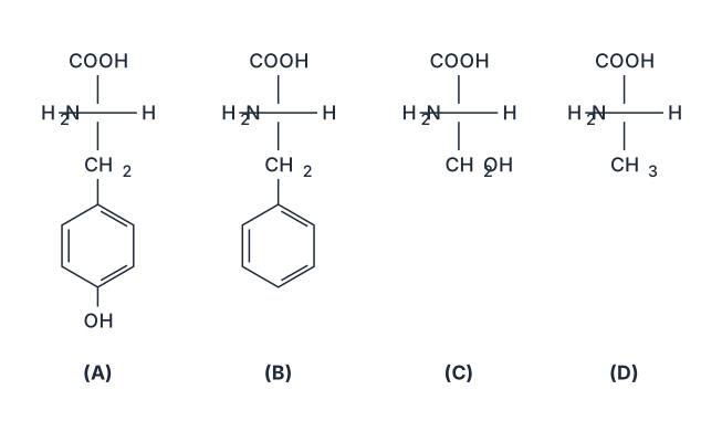
›Reveal solutionSolution
The amino acid classically obtained from cheese is tyrosine (Greek tyros = cheese), whose side chain is a p-hydroxybenzyl group. That is the structure in option (A).
The concept first: naming amino acids by their source
Many amino acids carry the fingerprint of the material they were first isolated from, and this makes an easy memory hook:
- Tyrosine — from Greek tyros, "cheese"; Justus von Liebig obtained it in 1846 by alkaline hydrolysis of casein, the chief protein of cheese.
- Glycine — from glykys, "sweet" (it tastes sweet).
- Asparagine — first found in asparagus juice.
- Cysteine — first isolated from urinary cystine stones.
- Serine — from sericum, silk (silk fibroin is rich in it).
Step-by-step: identify each structure
All four options are drawn as Fischer-type formulae with COOH on top, H2N to the left, H to the right and the side chain (R group) below. So the whole question reduces to reading the R group:
- (A) R =−CH2−C6H4−OH (benzene ring bearing OH at the para position) ⇒ HO−C6H4−CH2−CH(NH2)−COOH = tyrosine. This is the aromatic, phenolic amino acid — its −OH makes it weakly acidic and lets it be phosphorylated in signalling proteins. …
- TG EAPCET 2024Set ap-2024-05-07-FN1 markMCQQ.Phosphodiester linkage that joins nucleotides together is present between which carbons of pentose sugar? (A) 3′,3′ (B) 5′,3′ (C) 5′,2′ (D) 3′,2′
›Reveal solutionSolution
The phosphodiester bond links the 5' carbon of one nucleotide's sugar to the 3' carbon of the next nucleotide's sugar, forming the sugar-phosphate backbone. The correct answer is (B).
Concept & Intuition
Nucleotides are the building blocks of nucleic acids (DNA and RNA). Each nucleotide has three parts: a nitrogenous base, a pentose sugar (ribose or deoxyribose), and a phosphate group. When nucleotides join to form a chain, a phosphodiester linkage is created — this is a covalent bond where a phosphate group bridges two sugar molecules. The key question is: which carbons on the sugars are involved?
Think of the sugar as having numbered carbons (1' to 5'). The phosphate group attaches to the 5' carbon of one nucleotide's sugar and to the 3' carbon of the next nucleotide's sugar. This creates a directional backbone (5' → 3'), which is fundamental to DNA replication, transcription, and the overall structure of nucleic acids.
Step-by-Step Reasoning
-
Identify the sugar carbons
In a pentose sugar (ribose or deoxyribose), the carbons are numbered 1' through 5'. The base attaches at the 1' carbon. The 5' carbon is part of a CH₂OH group, and the 3' carbon has a hydroxyl (OH) group (in a free nucleotide).
-
Understand how nucleotides link
When two nucleotides join, the phosphate group on the 5' carbon of one nucleotide reacts with the hydroxyl group on the 3' carbon of the next nucleotide. This forms a phosphodiester bond — a phosphate diester bridge between the two sugars.
-
Confirm the specific carbons
The bond is always between the 5' carbon of one sugar and the 3' carbon of the adjacent sugar. This is a universal feature of DNA and RNA strands. …
-
- TG EAPCET 2024Set eng-2024-05-10-AN1 markMCQQ.The effects that aspirin can produce in the body are (A) A, B, C (B) A, C, D (C) A, B, E (D) C, D, E
›Reveal solutionSolution
Aspirin’s effects stem from its ability to inhibit prostaglandin synthesis — this leads to pain relief (analgesic), fever reduction (antipyretic), and anti-inflammatory action, but also to increased bleeding tendency (not a blood-clotting effect). The correct combination is A, C, D.
Aspirin (acetylsalicylic acid) is one of the most widely used drugs in medicine. To understand which effects it actually produces, you need to connect its biochemical mechanism to its clinical outcomes. The key is that aspirin irreversibly inhibits the enzyme cyclooxygenase (COX), which is responsible for making prostaglandins. Prostaglandins are chemical messengers that cause pain, fever, and inflammation — but they also protect the stomach lining and help platelets clump together to form clots.
So when aspirin blocks prostaglandin production, it removes the signals for pain, fever, and inflammation. That’s why it works as an analgesic (painkiller), antipyretic (fever reducer), and anti-inflammatory agent. But the same blockade also removes the protective prostaglandins in the stomach (leading to ulcer risk) and prevents platelet aggregation — which increases bleeding time, not clotting. Aspirin does not promote blood clotting; it does the opposite.
Now let’s match this to the options given. The effects listed are typically:
- A: Analgesic (pain relief)
- B: Blood-clotting (promotes clotting)
- C: Antipyretic (fever reduction)
- D: Anti-inflammatory (reduces swelling)
- E: Ulcer formation in stomach (a side effect, not a therapeutic effect)
The question asks for the effects aspirin can produce in the body — this includes both intended therapeutic effects and notable side effects. Let’s go step by step.
-
Analgesic effect (A) — Aspirin is a classic analgesic, effective for mild to moderate pain like headaches, muscle aches, and arthritis. It works by reducing prostaglandins that sensitize nerve endings to pain. So A is correct.
-
Blood-clotting effect (B) — This is the classic trap. Aspirin is actually an antiplatelet drug — it prevents platelets from sticking together by blocking thromboxane A₂ (a prostaglandin derivative). This prolongs bleeding time and is used in low doses to prevent heart attacks and strokes. It does not promote clotting. So B is incorrect.
-
Antipyretic effect (C) — Fever is caused by prostaglandins acting on the hypothalamus. Aspirin lowers fever by inhibiting their synthesis there. So C is correct.
-
Anti-inflammatory effect (D) — At higher doses, aspirin reduces inflammation (swelling, redness, heat) by suppressing prostaglandins at the site of tissue injury. So D is correct. …
- TG EAPCET 2023Set ap-2023-05-10-AN1 markMCQQ.Match the following List – I (Vitamin) A) Thiamine B) Riboflavin C) Ascorbic acid D) Vitamin D List-II (Deficiency disease) I Scurvy II Xerophthalmia III Cheilosis IV Beri Beri V Rickets The correct answer is (A) A – IV, B – III, C – V, D – II (B) A – III, B – IV, C – II, D – V (C) A – IV, B – III, C – I, D – V (D) A – I, B – III, C – II, D – IV
›Reveal solutionSolution
Match each vitamin to its classic deficiency disease by recalling their biochemical roles: Thiamine → Beri Beri, Riboflavin → Cheilosis, Ascorbic acid → Scurvy, Vitamin D → Rickets. The correct answer is (C).
Vitamins are organic micronutrients essential for specific metabolic functions, and their absence produces characteristic deficiency diseases. The key is to connect each vitamin's biochemical role to the pathology it prevents.
Thiamine (Vitamin B₁) is a coenzyme in carbohydrate metabolism, particularly in the decarboxylation of pyruvate and α-ketoglutarate. Without it, energy production falters, especially in high-demand tissues like nerves and heart. The result is Beri Beri, which presents as peripheral neuropathy (dry beri beri) or cardiac failure (wet beri beri). So A → IV.
Riboflavin (Vitamin B₂) forms the prosthetic groups FAD and FMN, critical in redox reactions. Deficiency affects rapidly dividing tissues—skin and mucous membranes. The hallmark is Cheilosis (painful cracks at the corners of the mouth), often accompanied by glossitis and dermatitis. Thus B → III.
Ascorbic acid (Vitamin C) is the cofactor for prolyl and lysyl hydroxylases, enzymes that stabilize collagen by hydroxylating proline and lysine residues. Without it, collagen becomes structurally weak, leading to Scurvy: bleeding gums, poor wound healing, petechiae, and bone fragility. Hence C → I. …
- TG EAPCET 2022Set ap-2022-07-30-FN1 markMCQQ.The correct statement for the formation of nitronium ion intermediate for an electrophilic aromatic nitration is (A) Both H2SO4 and HNO3 serve as acids (B) H2SO4 serves as acid and HNO3 as base (C) H2SO4 serves as base and HNO3 as acid (D) Both H2SO4 and HNO3 serve as bases
›Reveal solutionSolution
In the classic nitration of benzene, concentrated sulfuric acid protonates nitric acid, forcing it to lose water and form the electrophilic nitronium ion (NO2+). Here H2SO4 acts as the acid (proton donor) and HNO3 acts as the base (proton acceptor). The correct choice is (B).
The key to this question is understanding the Brønsted–Lowry acid–base roles in the formation of the nitronium ion. In electrophilic aromatic nitration, we need a powerful electrophile — the nitronium ion NO2+ — which is generated in situ from nitric acid. Concentrated sulfuric acid is not just a solvent; it plays an active chemical role.
-
Identify the goal: We want to convert HNO3 into NO2+. Nitric acid alone is not acidic enough to lose OH− (as water) on its own. It needs a stronger acid to protonate one of its oxygen atoms, making water a better leaving group.
-
Recognize the stronger acid: Sulfuric acid (H2SO4) is a much stronger acid than nitric acid (HNO3). In a mixture of the two, H2SO4 will donate a proton (H+) to HNO3.
-
Write the first step — proton transfer:
HNO3 accepts a proton from H2SO4:
HNO3+H2SO4⇌H2NO3++HSO4−
Here, H2SO4 is the acid (proton donor) and HNO3 is the base (proton acceptor).
- Write the second step — loss of water: The protonated nitric acid (H2NO3+) is unstable and readily loses a water molecule: H2NO3+→NO2++H2O …
-
- TG EAPCET 2021Set ap-2021-08-09-AN1 markMCQQ.The Lewis acid in the following is (A) PH3 (B) H2S (C) C2H4 (D) B2H6
›Reveal solutionSolution
A Lewis acid is an electron‑pair acceptor. Among the given molecules, only diborane (B2H6) has an empty orbital that can accept a pair of electrons, making it the Lewis acid. The correct option is (D).
Concept & Intuition
Lewis acids are species that can accept an electron pair. They typically have an incomplete octet, a vacant low‑energy orbital, or a positive charge. Lewis bases are electron‑pair donors. To identify the Lewis acid among the options, we check each molecule’s electronic structure: does it have a site that wants electrons?
- PH3 (phosphine) – Phosphorus has a lone pair and a complete octet. It donates electrons, so it acts as a Lewis base, not an acid.
- H2S (hydrogen sulfide) – Sulfur has two lone pairs and a full octet. It is an electron‑pair donor (Lewis base).
- C2H4 (ethene) – The carbon atoms are sp2‑hybridized with a full octet and a π‑bond. The π‑electrons are available for donation (e.g., to a metal), so ethene is a Lewis base. …
- TG EAPCET 2021Set ap-2021-08-09-FN1 markMCQQ.The structure of β-D-2-deoxyribose is (A) [FIGURE] Haworth furanose ring: HOH2C at the top-left and ring O at the top, OH at the top-right (anomeric C); ring interior labelled H, H; H on the left; H on the right; bottom substituents H and OH (B) [FIGURE] Haworth furanose ring: HOH2C at the top-left and ring O at the top, H at the top-right (anomeric C); ring interior labelled H, H; H on the left; OH on the right; bottom substituents OH and H (C) [FIGURE] Haworth furanose ring: HOH2C at the top-left and ring O at the top, OH at the top-right (anomeric C); ring interior labelled H, OH; H on the left; H on the right; bottom substituents H and H (D) [FIGURE] Haworth furanose ring: HOH2C at the top-left and ring O at the top, OH at the top-right (anomeric C); ring interior labelled H, H; H on the left; H on the right; bottom substituents OH and H
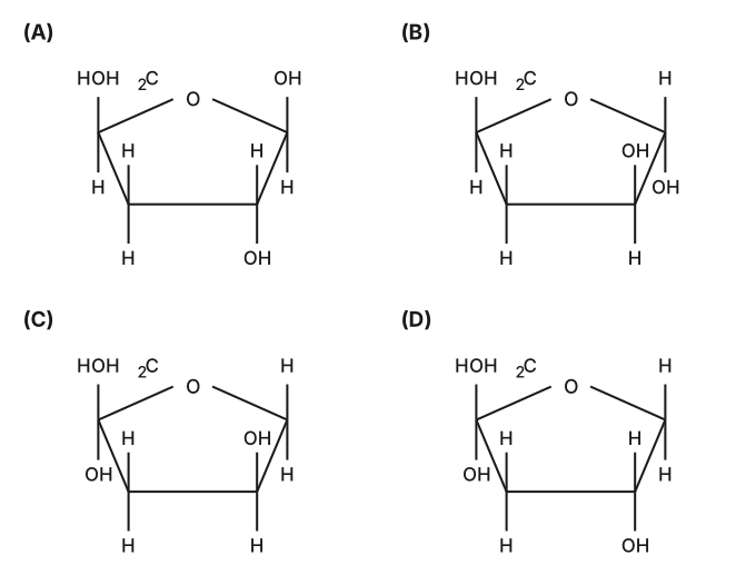
›Reveal solutionSolution
β-D-2-deoxyribose is a five-membered furanose ring with the anomeric OH up (β), no hydroxyl at C-2 (deoxy), and the standard D-configuration at C-3 and C-4. The answer is (D).
The name "β-D-2-deoxyribose" encodes three pieces of structural information that we need to decode systematically.
What "2-deoxy" means: The prefix tells us that carbon-2 (the first carbon after the anomeric carbon in the ring) lacks a hydroxyl group. Instead of OH, it carries only hydrogen. This is the defining feature of deoxyribose versus ribose.
What "D" means: The D/L designation refers to the configuration at the highest-numbered chiral center in the Fischer projection—for a pentose, that's C-4 (the carbon bearing the CH2OH group in the ring form). In the Haworth projection of a D-sugar, the CH2OH group projects upward (above the plane of the ring).
What "β" means: The β-anomer has the anomeric hydroxyl (at C-1) on the same side as the CH2OH group. Since we've established that CH2OH points up in D-sugars, the anomeric OH must also point up.
Now let's build the structure step by step:
-
Start with the furanose ring skeleton. All options show a five-membered ring with oxygen at the top-right position and CH2OH at the top-left—this is the standard Haworth orientation for a D-pentose.
-
Place the anomeric hydroxyl (C-1). The β-configuration requires OH pointing up at the anomeric carbon (top-right position). This eliminates option (B), which has H pointing up at this position.
-
Identify C-2 (the "deoxy" position). Moving clockwise from the anomeric carbon, C-2 is the next ring carbon. The "2-deoxy" designation means this carbon has no OH group—only hydrogens. In the Haworth projection, both the substituent on the ring carbon and the interior label at this position should be H, not OH. This eliminates option (C), which shows OH in the ring interior at C-2. …
-
🎓Unlock everything free for 14 days
- ✓Full step-by-step solutions
- ✓Concept-first explanations
- ✓Methods, shortcuts & mistakes
- ✓PYQ mapping + timed mock tests
Full access for 14 days. No credit card required.