Human Dentition – A First Look
Think about what your teeth actually do. You don't just bite and swallow. You tear, you crush, you grind. A piece of meat needs a different kind of treatment than a carrot or a nut. Your mouth is a processing plant, and each tooth is a specialised tool for a specific job.
That's the whole idea behind human dentition: different teeth for different tasks, arranged in a precise pattern that appears twice in a lifetime.
The Three Big Words
Before we get to the teeth themselves, you need three terms that describe the arrangement and replacement of teeth. They sound fancy, but they're simple.
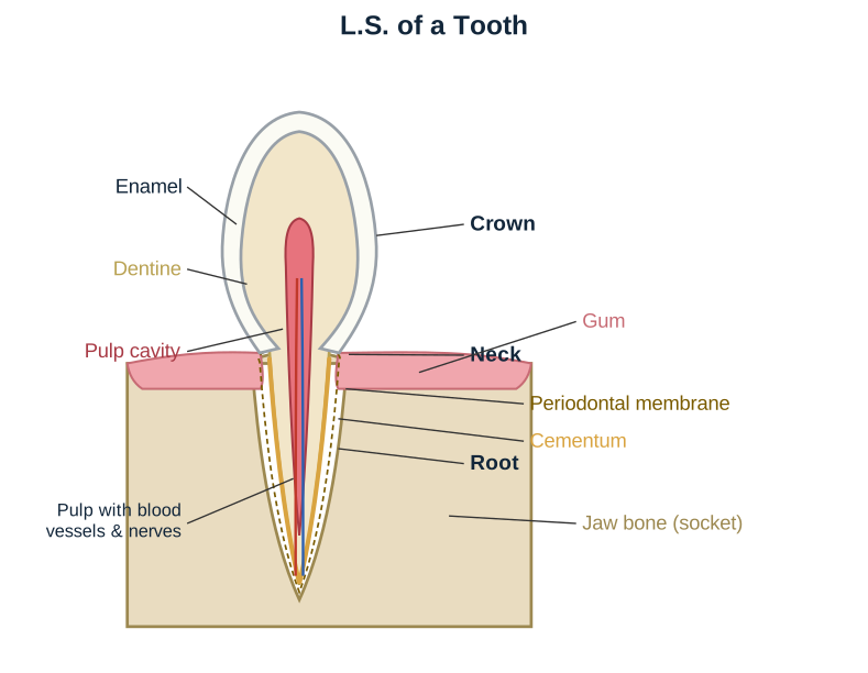
Thecodont – each tooth sits in its own socket (a deep bony pit called an alveolus) inside the jawbone. This is what gives your teeth their firm anchorage. Almost all mammals, including humans, are thecodont. Crocodiles are another example. Reptiles like snakes are not – their teeth are fused to the bone.
Diphyodont – you get two sets of teeth in your life. The first set is the milk teeth (deciduous teeth), which start falling out around age 6. The second set is the permanent teeth. "Di" = two, "phy" = growth. Two eruptions.
Heterodont – your teeth are not all the same shape. They are differentiated into four types: incisors, canines, premolars, and molars. "Hetero" = different, "odont" = tooth. Compare this to a shark, which has rows of identical teeth (homodont).
Human dentition is thecodont, diphyodont, and heterodont. This is a standard exam fact. Memorise the three terms together.
The Four Tooth Types
Here is what each type does, and where it sits in the mouth.
| Tooth Type | Number (per half jaw) | Primary Job | Shape |
|---|
| Incisors | 2 | Cutting and shearing food (like a chisel) | Flat, sharp-edged crown |
| Canines | 1 | Tearing and piercing (especially tough food) | Pointed, conical crown |
| Premolars | 2 | Crushing and grinding (transitional) | Two cusps (bicuspid) |
| Molars | 3 | Heavy grinding and chewing | Large, multi-cusped surface |
The numbers above are for one half of one jaw (upper or lower). So in the upper jaw, you have 2 incisors, 1 canine, 2 premolars, and 3 molars on the right side – and the same on the left. That gives 8 teeth per half jaw, 16 per jaw, 32 total in a full permanent set.
The Dental Formula: 2123 / 2123
This is the shorthand that summarises the entire arrangement. It is written as a fraction, but it is not a mathematical fraction. It is a code.
Upper jaw (maxilla): 2 incisors, 1 canine, 2 premolars, 3 molars
Lower jaw (mandible): 2 incisors, 1 canine, 2 premolars, 3 molars
So the formula is:
2 1 2 32 1 2 3
This counts one side only (the right half of the upper jaw over the right half of the lower jaw). To get the total number of teeth, multiply each number by 2 (for left and right) and then add upper and lower. …