Q.(a)
🔒You're viewing a preview — the full solution, concept, methods & PYQ mapping are locked.
🔒 Start your 14-day free trial to unlock the full solution →Part (a)Concept understanding — Mendelian Genetics Basics
Mendelian Genetics Basics
Imagine you have a box of coloured beads — red and white. If you pick one bead from the box, you get either red or white. Now imagine that the colour of your eyes, or the shape of your earlobe, is decided by something like that: a tiny "packet" inside your cells that comes in two versions, and you inherit one from each parent. That is the core idea of Mendelian genetics.
The everyday intuition
You have probably noticed that children often look like their parents — same hair colour, same dimples, same height. But they are never exact copies. Why? Because each parent contributes half of the "instructions" for building a child. Those instructions come in pairs, one from mother and one from father. Sometimes one instruction overrides the other; sometimes they blend. Gregor Mendel, a 19th-century monk, figured out the rules by watching pea plants — tall vs short, yellow vs green seeds — and counting what appeared in the next generation.
The precise meaning
Mendelian genetics is the study of how traits are passed from parents to offspring through genes. A gene is a unit of heredity — a stretch of DNA that codes for a specific characteristic, like flower colour. Each gene comes in different versions called alleles. For every gene, you inherit two alleles: one from your mother, one from your father.
If the two alleles are identical, you are homozygous for that trait. If they are different, you are heterozygous. In a heterozygous pair, one allele may be dominant — it shows up in the appearance — and the other recessive — it stays hidden unless both alleles are recessive.
Mendel's key insight was that traits are not blended like paint. Instead, alleles remain separate and are passed on intact. A recessive allele can skip a generation and reappear later, unchanged.
Why it matters
Mendelian genetics is the foundation of modern biology. It explains:
- Why some diseases run in families (like cystic fibrosis or sickle-cell anaemia)
- How plant and animal breeders create new varieties
- Why you might have your grandmother's eyes but not your mother's
The NCERT textbook states that Mendel's work established the laws of inheritance — the Law of Dominance, the Law of Segregation, and the Law of Independent Assortment. These laws describe how alleles separate during the formation of eggs and sperm, and how different genes are inherited independently of one another.
Key terms at a glance
- Gene: a unit of heredity on a chromosome
- Allele: a variant form of a gene
- Dominant: the allele that expresses itself even when paired with a different allele
- Recessive: the allele that expresses itself only when paired with an identical recessive allele
- Homozygous: having two identical alleles for a gene
- Heterozygous: having two different alleles for a gene …
Part (b)Concept understanding — Lac Operon Catabolite Repression
Imagine you are a factory manager. You have two raw materials: a high-grade fuel that your machines run on perfectly, and a low-grade backup fuel that works but is harder to use. As long as the good fuel is available, you would never waste time and energy switching to the backup. But if the good fuel runs out, you immediately switch to the backup to keep production going.
That is exactly what catabolite repression does inside a bacterium like E. coli. It is the cell's way of saying: "Use the best fuel first; don't bother with the second-best until you absolutely have to."
The Two Fuels: Glucose and Lactose
E. coli bacteria love glucose. It is their favourite energy source — easy to break down, gives quick energy. Lactose (milk sugar) is harder to digest; the cell needs to build special enzymes (like β-galactosidase) to break it down. These enzymes are coded by the lac operon.
The cell has a simple rule: If glucose is present, do not waste energy making lactose-digesting enzymes. That is catabolite repression. It is a global regulatory mechanism that ensures glucose is used first, even when lactose is also available.
Catabolite repression is sometimes called the glucose effect. It is not unique to the lac operon — it affects many operons that break down alternative sugars. But the lac operon is the classic textbook example.
How It Works: The Molecular Switch
The key player is a molecule called cAMP (cyclic AMP). Its level inside the cell is inversely related to glucose concentration:
- When glucose is high: cAMP levels are low.
- When glucose is low: cAMP levels rise.
cAMP binds to a protein called CAP (Catabolite Activator Protein). The cAMP–CAP complex then binds to a specific site near the lac operon's promoter. This binding dramatically increases the rate of transcription — it is like pressing the accelerator pedal.
So here is the logic:
- Glucose present (high): Low cAMP → CAP cannot bind → lac operon is barely transcribed, even if lactose is around. The cell ignores lactose.
- Glucose absent (low): High cAMP → CAP binds → lac operon is fully activated. Now, if lactose is also present, the operon switches on fully and the cell digests lactose.
Catabolite repression is a positive control mechanism. The CAP–cAMP complex activates transcription. This is different from the lac repressor, which blocks transcription when lactose is absent. The lac operon is controlled by two switches: a negative one (repressor) and a positive one (CAP–cAMP). Both must be in the "on" position for maximum expression.
Why It Matters (Exam Perspective)
The NCERT textbook (Class 12 Biology, Chapter 6) presents catabolite repression as a fine-tuning mechanism. It explains that even when the lac repressor is removed (by lactose binding), transcription is still low unless glucose is absent. The CAP–cAMP complex is the "second key" that unlocks full expression.
Key points to remember for exams: …
Part (a)
(i) Pea (violet/white) vs Snapdragon (red/white):
| Feature | Garden pea | Snapdragon |
|---|---|---|
| F1 phenotype | All violet | All pink |
| F2 phenotype | 3 violet : 1 white | 1 red : 2 pink : 1 white |
| F2 genotype | 1 VV : 2 Vv : 1 vv | 1 RR : 2 Rr : 1 rr |
| Conclusion | Complete dominance | Incomplete dominance |
- Pea shows complete dominance (F1 violet, F2 3:1); snapdragon shows incomplete dominance (F1 pink, F2 1:2:1); ABO shows multiple alleles + codominance.
- In the switched-on lac operon lactose inactivates the repressor so RNA polymerase transcribes z,y,a; it is negative regulation because a repressor inhibits transcription.
Part (a)
(i) Comparison of flower-colour inheritance
In the garden pea, let V = violet (dominant) and v = white (recessive). Crossing true-breeding VV × vv:
- F1: all Vv → all violet (the dominant allele completely masks the recessive one).
- F2 (on selfing Vv × Vv): genotype 1 VV : 2 Vv : 1 vv; phenotype 3 violet : 1 white.
In the snapdragon (Antirrhinum), let R = red and r (or W) = white. Crossing RR × rr:
- F1: all Rr → all pink — an intermediate phenotype, because neither allele is completely dominant.
- F2: genotype 1 RR : 2 Rr : 1 rr; phenotype 1 red : 2 pink : 1 white.
| Feature | Garden pea (violet/white) | Snapdragon (red/white) |
|---|---|---|
| F1 phenotypic expression | All violet | All pink (intermediate) |
| F2 phenotypic ratio | 3 violet : 1 white | 1 red : 2 pink : 1 white |
| F2 genotypic ratio | 1 VV : 2 Vv : 1 vv | 1 RR : 2 Rr : 1 rr |
| Conclusion | Complete dominance | Incomplete dominance |
Conclusion: in the pea the heterozygote looks like the dominant parent, so the phenotypic and genotypic ratios differ (3:1 vs 1:2:1) — complete dominance. In the snapdragon the heterozygote is intermediate, so the phenotypic ratio equals the genotypic ratio (1:2:1) — incomplete dominance.
(ii) Two characteristics of ABO blood-group inheritance
- Multiple alleles: three alleles — I^A, I^B and i — control the trait (though any individual carries only two). …
Showing the 12 most recent of 19 on this concept.
- TG EAPCET 2026Set ap-2026-05-04-AN1 markMCQQ.Match the following List-1 List-2 A. linkage I. Common in cancer cells B. Recombination II. Parental gene combination C. Point mutation III. Non parental combination D. Chromosomal aberration IV. Sickle cell anemia V. Multiple phenotypes The correct answer is (A) A-II, B-V, C-IV, D-I (B) A-II, B-III, C-IV, D-I (C) A-I, B-IV, C-III, D-V (D) A-II, B-III, C-IV, D-V
›Reveal solutionSolution
This question tests your understanding of key genetic terms and their real-world examples. The correct mapping is A-II, B-III, C-IV, D-I, which corresponds to option (B).
Let’s build the intuition first. Each term in List-1 describes a specific genetic phenomenon, and List-2 gives either a cause, a consequence, or a classic example. The trick is to match each term with its most direct and well-known association — not just any possible connection, but the one that is textbook-standard for Indian exams.
-
A. Linkage → II. Parental gene combination
Linkage means genes located close together on the same chromosome tend to be inherited as a block. They do not assort independently, so the offspring often get the same combination of alleles that the parents had — hence “parental gene combination.” This is the classic definition: linkage preserves parental combinations unless crossing over breaks them.
-
B. Recombination → III. Non-parental combination
Recombination is the process that shuffles alleles between homologous chromosomes during meiosis (crossing over). The result is new combinations of genes that were not present together in either parent — these are called “non-parental” or “recombinant” combinations. So B matches III directly.
-
C. Point mutation → IV. Sickle cell anemia
A point mutation is a change in a single nucleotide base pair. Sickle cell anemia is the classic example: a single base substitution in the beta-globin gene (GAG → GTG) changes glutamic acid to valine, causing the disease. This is the go-to example in every syllabus.
-
D. Chromosomal aberration → I. Common in cancer cells …
-
- TG EAPCET 2026Set ap-2026-05-04-AN1 markMCQQ.Study the following and choose the correct statements I. If a single gene influences more than one phenotypic trait, it is called pleiotropy II. Due to absence of antibodies anti A and anti B, the persons with AB blood group are described as universal recipients III. Inheritance of sex linked dominant traits follow cris cross inheritance IV. If sex index ratio is 0.33, then sexual phenotype of Drosophila is intersex (A) I, III (B) II, IV (C) III, IV (D) I, II
›Reveal solutionSolution
Statements I and II are correct; III and IV are wrong, so the correct set is I, II — option (D).
I – Correct. When a single gene affects several phenotypic traits, the effect is called pleiotropy (e.g. the sickle-cell gene affects RBC shape, anaemia and malaria resistance). This is the standard definition.
II – Correct. Blood group AB carries both A and B antigens but no anti-A or anti-B antibodies in the plasma, so such persons can receive blood of any ABO type without agglutination — they are universal recipients.
III – Incorrect. Criss-cross inheritance (father → daughter → grandson) is the pattern of X-linked recessive traits. X-linked dominant traits do not show criss-cross inheritance — an affected father passes the trait to all his daughters but to none of his sons. …
- TG EAPCET 2026Set ap-2026-05-05-FN1 markMCQQ.Gene expression in eukaryotes could be regulated at I. Transcription level II. Translation level III. Splicing processing level IV. mRNA transport level from nucleus to the cytoplasm (A) I and II only (B) II, III and IV only (C) I, II and IV only (D) I, II, III and IV
›Reveal solutionSolution
Gene expression in eukaryotes can be regulated at every step from DNA to functional protein — transcription, splicing, mRNA transport, and translation are all control points. The correct answer is that all four levels are involved.
The key idea is that eukaryotic gene expression is not a simple, one-step process. Unlike prokaryotes, where transcription and translation happen in the same compartment, eukaryotes have a nucleus that separates these processes. This physical separation creates multiple opportunities for regulation — each step from DNA to a functional protein can be turned up, turned down, or fine-tuned.
Let’s walk through each level mentioned in the question.
-
Transcription level — This is the most fundamental control point. Before any protein can be made, the gene must be transcribed into RNA. In eukaryotes, transcription is regulated by transcription factors binding to promoter and enhancer regions, chromatin remodeling (opening or closing DNA), and epigenetic modifications like DNA methylation. This is the primary level where cells decide whether a gene is "on" or "off."
-
Splicing processing level — After transcription, the pre-mRNA undergoes splicing to remove introns and join exons. But here’s the twist: alternative splicing allows a single gene to produce multiple different mRNA variants by choosing different combinations of exons. This is a major regulatory mechanism — the same gene can code for different proteins in different tissues or at different times, all controlled at the splicing stage.
-
mRNA transport level from nucleus to cytoplasm — Once the mature mRNA is ready, it must be exported through nuclear pores into the cytoplasm. This transport is not automatic; it is regulated. Some mRNAs are held back in the nucleus until a specific signal arrives, or their export efficiency is controlled. If an mRNA never reaches the cytoplasm, it cannot be translated — so this is a genuine regulatory step. …
-
- TG EAPCET 2025Set ap-2025-04-29-AN1 markMCQQ.The disease sickle cell anemia is caused by substitution of ‘A’ by ‘B’ of ‘C’ globin chain of hemoglobin molecule. Identify A, B and C respectively (A) A – Alanine, B – Glutamic acid, C – Beta (B) A – Glutamic acid, B – Valine, C – Beta (C) A – Valine, B – Glutamic acid, C – Alpha (D) A – Valine, B – Serine, C – Beta
›Reveal solutionSolution
Sickle cell anemia results from a point mutation where glutamic acid is replaced by valine at the sixth position of the beta-globin chain of hemoglobin. The correct option is (B).
Sickle cell anemia is a classic example of a genetic disorder caused by a single point mutation, leading to a change in a single amino acid in a protein. This seemingly small change has profound effects on the structure and function of hemoglobin, ultimately altering the shape of red blood cells and causing the disease's characteristic symptoms. Understanding this specific molecular change is key to grasping the pathology of sickle cell anemia.
Here's a breakdown of the molecular basis of sickle cell anemia:
-
Hemoglobin Structure: Hemoglobin is the oxygen-carrying protein in red blood cells. In adults, it is primarily composed of four polypeptide chains: two alpha (α) globin chains and two beta (β) globin chains. Each chain contains a heme group that binds oxygen.
-
The Genetic Defect: Sickle cell anemia arises from a mutation in the gene that codes for the beta-globin chain. This is a point mutation, meaning a change in a single nucleotide base pair in the DNA sequence.
-
Identifying the Globin Chain (C): The specific mutation responsible for sickle cell anemia occurs in the beta (β) globin chain. This is why the disease is often referred to as a "beta-thalassemia" in some contexts, though sickle cell anemia has its own distinct pathology.
-
Identifying the Amino Acid Substitution (A and B):
- At the sixth position of the beta-globin chain, the normal amino acid is glutamic acid (A). Glutamic acid is a hydrophilic (water-loving) amino acid.
- Due to the point mutation (a change from GAG to GTG in the mRNA codon), valine (B) is substituted in place of glutamic acid at this sixth position. Valine is a hydrophobic (water-fearing) amino acid. …
-
- TG EAPCET 2025Set ap-2025-04-29-AN1 markMCQQ.Study the following and identify the correct combinations(A) I & III (B) II & III (C) II & IV (D) I & II
Phenomenon Phenotypic ratio Genotypic ratio I Co-dominance 1:2:1 1:2:1 II Incomplete dominance 3:1 1:2:1 III Monohybrid test cross 1:1 1:1 IV Dihybrid test cross 9:3:3:1 1:1:1:1 ›Reveal solutionSolution
Only I (co-dominance: 1:2:1 and 1:2:1) and III (monohybrid test cross: 1:1 and 1:1) are stated correctly. Incomplete dominance gives a 1:2:1 phenotypic ratio (not 3:1), and a dihybrid test cross gives 1:1:1:1 phenotypes (not 9:3:3:1). Hence the correct combination is I & III — option (A).
The concept first
Two different things are being asked in every row:
- The genotypic ratio comes only from which gametes combine — it is pure Punnett-square bookkeeping and is the same whatever the dominance relationship is.
- The phenotypic ratio depends on how the alleles interact:
- Complete dominance — the heterozygote looks like the dominant homozygote, so two genotype classes fuse and 1:2:1 collapses to 3:1.
- Incomplete dominance — the heterozygote is intermediate (e.g. pink Mirabilis), so nothing fuses and the phenotypic ratio equals the genotypic ratio, 1:2:1.
- Co-dominance — the heterozygote shows both phenotypes together (e.g. blood group AB), so again nothing fuses and the phenotypic ratio equals the genotypic ratio, 1:2:1.
So the memorable rule is: in both incomplete dominance and co-dominance, phenotypic ratio = genotypic ratio =1:2:1.
Step-by-step
Step 1 — Row I: Co-dominance.
Cross IAIB×IAIB (or any co-dominant heterozygote × itself). Gametes from each parent: 21IA, 21IB.
Offspring=41IAIA:21IAIB:41IBIB=1:2:1 (genotypic)
Because the heterozygote expresses both alleles, it is its own distinct phenotype, so the phenotypic ratio is also 1:2:1. The row says 1:2:1 and 1:2:1 — correct.
Step 2 — Row II: Incomplete dominance.
Rr×Rr gives genotypes RR:Rr:rr=1:2:1 — the row's genotypic ratio is right. But the heterozygote Rr is pink, distinct from red RR and white rr, so the phenotypes are 1 red :2 pink :1 white =1:2:1. The row prints 3:1, which is the complete-dominance result. Row II is incorrect.
Step 3 — Row III: Monohybrid test cross.
A test cross is heterozygote × recessive homozygote: Aa×aa. Gametes: 21A,21a from one side; only a from the other.
Offspring=21Aa:21aa=1:1 …
- TG EAPCET 2025Set ap-2025-04-29-FN1 markMCQQ.Study the following and identify the correct statements: I. The alleles IA and IB regarding blood groups are dominant over IO, and IA and IB are co-dominant II. In some fishes, reptiles and birds, females are heterogametic (ZW) and males are homogametic (ZZ). III. Genic balance theory states that Y-chromosome is essential for determination of sex in male Drosophila IV. Person with O blood group are called universal donors because their RBC contain both antigens A and B. (A) I, II (B) III, IV (C) I, III (D) II, IV
›Reveal solutionSolution
The question tests four statements on genetics — blood group inheritance, sex determination in ZW systems, the genic balance theory in Drosophila, and the basis of universal donation. Statements I and II are correct; III and IV are false. The correct option is (A).
Let’s examine each statement carefully, one by one.
-
Statement I: The alleles IA and IB regarding blood groups are dominant over IO, and IA and IB are co-dominant.
This is textbook ABO blood group genetics. The IA and IB alleles each produce a specific antigen (A and B respectively), while IO produces no functional antigen. Both IA and IB are dominant over IO, so a person with genotype IAIO has blood group A, and IBIO gives group B. When both IA and IB are present together, both antigens are expressed equally — that is co-dominance, giving blood group AB.
Statement I is correct.
-
Statement II: In some fishes, reptiles and birds, females are heterogametic (ZW) and males are homogametic (ZZ).
In the ZW sex-determination system, the female has two different sex chromosomes (ZW) and the male has two identical ones (ZZ). This is indeed found in birds, many reptiles, and some fishes. The opposite (XX/XY) system is typical in mammals and Drosophila.
Statement II is correct.
-
Statement III: Genic balance theory states that Y-chromosome is essential for determination of sex in male Drosophila.
This is a classic trap. In Drosophila melanogaster, sex is determined not by the presence of a Y chromosome, but by the ratio of X chromosomes to autosomes (the X:A ratio). The Y chromosome in Drosophila is required only for male fertility, not for male determination. The genic balance theory, proposed by Calvin Bridges, explicitly states that the Y chromosome is not essential for maleness — a fly with XXY is female, and a fly with XO (no Y) is male but sterile.
Statement III is false. …
-
- TG EAPCET 2025Set ap-2025-04-30-FN1 markMCQQ.Consider the following statements Assertion (A): Skin colour in human beings is a polygenetic trait. Reason (R): Human skin colour is controlled by cumulative effect of genes. The correct answer is (A) Both (A) and (R) are true, (R) is the correct explanation of (A) (B) Both (A) and (R) are true, (R) is not the correct explanation of (A) (C) (A) is true, but (R) is false (D) (A) is false, but (R) is true
›Reveal solutionSolution
Skin colour is polygenic because multiple genes act cumulatively/additively; R explains A.
Assertion: Skin colour in human beings is a polygenic trait - a phenotype governed by more than one gene, showing a continuous gradation of expression rather than discrete classes. …
- TG EAPCET 2024Set ap-2024-05-07-AN1 markMCQQ.Observe the phenotypic ratios given below A. 9:3:3:1 B. 1:2:1 C. 1:1 Arrange the ratios in the order of monohybrid test cross; incomplete dominance; and dihybrid cross (A) B-C-A (B) C-B-A (C) B-A-C (D) A-B-C
›Reveal solutionSolution
A monohybrid test cross gives 1:1 (ratio C), incomplete dominance gives 1:2:1 (ratio B) and a dihybrid cross gives 9:3:3:1 (ratio A). In the order asked, that is C – B – A, i.e. option (B).
The concept first
Ratios in genetics are not things to be memorised as trivia — each one is generated by a specific set of gametes, and if you can build the Punnett square you never have to remember anything.
Two questions decide every ratio:
- How many gene pairs are segregating? One → monohybrid arithmetic. Two → multiply the two monohybrid outcomes (Law of Independent Assortment).
- Is the heterozygote phenotypically identical to the dominant homozygote, or does it look different? If identical → complete dominance, and genotypes collapse into fewer phenotypes. If different → incomplete dominance, and no collapsing happens.
Step-by-step
- Monohybrid test cross → ratio C (1:1). A test cross means crossing an individual of unknown genotype with the homozygous recessive. For a heterozygote:
Tt×tt
Gametes: T and t from the first parent; only t from the second.
Offspring: 21Tt (tall) and 21tt (dwarf).
∴phenotypic ratio=1:1⇒C
This is exactly why a test cross works — a heterozygote betrays itself by producing recessive offspring.
- Incomplete dominance → ratio B (1:2:1). In Mirabilis jalapa (four o'clock plant), RR = red, rr = white, and Rr is pink — its own distinct phenotype. Selfing the F1:
Rr×Rr→1RR:2Rr:1rr=1 red:2 pink:1 white
Because the heterozygote is visibly different, the 2Rr class does not merge with the 1RR class. So the phenotypic ratio equals the genotypic ratio:
1:2:1⇒B …
- TG EAPCET 2024Set ap-2024-05-07-AN1 markMCQQ.If karyotype of a Drosophila is AA-XXXY, its sexual phenotype is (A) Matafemale (B) Metamale (C) Intersex (D) Female with Y chrosome
›Reveal solutionSolution
In Drosophila, sex is determined by the ratio of X chromosomes to sets of autosomes (the sex index). For a karyotype of AA-XXXY, the sex index is 1.5, which results in a Metafemale phenotype.
The sexual phenotype of Drosophila is determined by a mechanism known as the genic balance theory, proposed by C.B. Bridges. Unlike humans where the presence of a Y chromosome primarily determines maleness, in Drosophila, the balance between the number of X chromosomes and the number of sets of autosomes dictates the sex. The Y chromosome in Drosophila is primarily involved in male fertility, not sex determination itself.
This balance is quantified by the "sex index," which is calculated as the ratio of the number of X chromosomes (X) to the number of sets of autosomes (A).
Sex Index (I)=Number of sets of autosomesNumber of X chromosomes
Different values of this index correspond to different sexual phenotypes:
- I=1.0: Normal Female
- I>1.0: Metafemale (also called Superfemale)
- I=0.5: Normal Male
- I<0.5: Metamale (also called Supermale)
- 0.5<I<1.0: Intersex
Let's apply this concept to the given karyotype.
-
Identify the number of X chromosomes: The given karyotype is AA-XXXY. Here, 'XXXY' indicates there are three X chromosomes. So, X=3.
-
Identify the number of sets of autosomes: The 'AA' in the karyotype represents two sets of autosomes. So, A=2. …
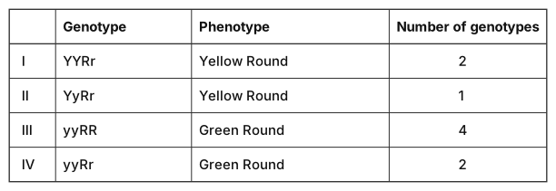
- TG EAPCET 2024Set ap-2024-05-07-FN1 markMCQQ.The table given below is related to checker board of dihybrid F2 progeny of Mendel’s experiment. Identify correct combinations Genotype | Phenotype | Number of genotypes I YYRr Yellow Round 2 II YyRr Yellow Round 1 III yyRR Green Round 4 IV yyRr Green Round 2 Options : (A) I and II (B) II and IV (C) III and IV (D) I and IV
›Reveal solutionSolution
In the 16-square dihybrid Punnett square, YYRr appears 2 times and yyRr appears 2 times (both correct), while YyRr appears 4 (not 1) and yyRR appears 1 (not 4). The correct combinations are I and IV — option (D).
The concept first
The dihybrid F2 is nothing more than two independent monohybrid crosses multiplied together — that is Mendel's Law of Independent Assortment in action. YyRr makes four gamete types, YR, Yr, yR, yr, in equal 1:1:1:1 proportion, so the checker board has 4×4=16 boxes.
Because the loci are independent, the number of boxes for any genotype is just the product of its two single-locus frequencies out of 4:
homozygote at a locus→1/4,heterozygote at a locus→2/4
So:
- homozygous at both loci → 1×1=1 box,
- heterozygous at one locus → 2×1=2 boxes,
- heterozygous at both loci → 2×2=4 boxes. That single rule answers the whole question without drawing anything.
Step-by-step
- Write the full F2 genotypic ratio (the 9 genotypes in 16 boxes):
1YYRR:2YYRr:2YyRR:4YyRr:1YYrr:2Yyrr:1yyRR:2yyRr:1yyrr
(These 16 collapse into the familiar phenotypic 9:3:3:1.) …
- TG EAPCET 2023Set ap-2023-05-10-FN1 markMCQQ.Study the following tables and match the correct combination for the dihybrid F2 progeny Genotype Phenotype Genotypes Number I YYRr Yellow round 2 II YyRr Yellow round 1 III yyRR Green round 4 IV yyRr Green round 2 (A) III and IV (B) II and IV (C) I and II (D) I and IV
›Reveal solutionSolution
In a dihybrid cross (YyRr × YyRr), the F2 progeny follow a 9:3:3:1 phenotypic ratio. The table lists genotypes and their counts; we must match each row to the correct number of individuals. The correct combination is I and IV.
The question tests your understanding of Mendelian dihybrid inheritance — specifically, the genotypic and phenotypic ratios that arise from a cross between two double heterozygotes (YyRr×YyRr). The key is to recall that each trait (seed colour and seed shape) segregates independently, giving a 9:3:3:1 phenotypic ratio in the F2 generation. But here, the table gives specific genotypes and asks you to verify the number of individuals with that genotype.
Let’s break it down step by step.
-
Set up the cross.
Both parents are YyRr (yellow, round). The gametes produced are YR, Yr, yR, yr in equal proportions (1:1:1:1). The Punnett square for a dihybrid cross has 16 equally likely combinations.
-
Determine the expected genotypic frequencies.
For any one gene pair, the monohybrid ratio is 1:2:1 (e.g., YY:Yy:yy). Since the two genes assort independently, the combined genotypic ratio is the product of the two monohybrid ratios.
- For colour: YY (1), Yy (2), yy (1).
- For shape: RR (1), Rr (2), rr (1). Multiply these to get the 16-cell Punnett square frequencies. For example, YYRR appears 1×1=1 time, YyRr appears 2×2=4 times, etc.
-
Match each row in the table to the expected count.
- Row I: YYRr — genotype YY (1) × Rr (2) = 2 individuals. The table says 2. Correct.
- Row II: YyRr — Yy (2) × Rr (2) = 4 individuals. The table says 1. Incorrect. …
-
- TG EAPCET 2023Set ap-2023-05-10-AN1 markMCQQ.Study the following and pick up the correct combinations S.No. Hormone Gland Effect of Hyper / Hyposecretion I Vasopressin Pituitary gland Diabetes insipidus II Calcitonin Parathyroid gland Cretinism III Cortisol Adrenal gland Addison's disease IV Insulin Pancreas Diabetes insipidus (A) I, II (B) III, IV (C) I, III (D) II, IV
›Reveal solutionSolution
The question tests your knowledge of hormones, their source glands, and the diseases caused by their abnormal secretion. Only combinations I (Vasopressin → Pituitary → Diabetes insipidus) and III (Cortisol → Adrenal → Addison's disease) are correct, so the answer is option (C).
The key here is to match each hormone with its correct gland and then with the specific disorder caused by either hypo- or hypersecretion. Many students mix up glands (like parathyroid vs. thyroid) or confuse diseases (like diabetes insipidus vs. mellitus). Let’s check each row carefully.
-
Row I: Vasopressin – Pituitary gland – Diabetes insipidus
Vasopressin (also called antidiuretic hormone, ADH) is secreted by the posterior pituitary. Its main job is to increase water reabsorption in the kidneys. When secretion is too low (hyposecretion), the kidneys cannot concentrate urine, leading to diabetes insipidus — excessive dilute urine and thirst. This match is correct.
-
Row II: Calcitonin – Parathyroid gland – Cretinism
Calcitonin is actually secreted by the thyroid gland (parafollicular cells), not the parathyroid. The parathyroid glands secrete parathyroid hormone (PTH). And cretinism is caused by hyposecretion of thyroid hormone (thyroxine) during childhood, not by calcitonin issues. So both the gland and the disease are wrong. This row is incorrect.
-
Row III: Cortisol – Adrenal gland – Addison's disease …
-
🎓Unlock everything free for 14 days
- ✓Full step-by-step solutions
- ✓Concept-first explanations
- ✓Methods, shortcuts & mistakes
- ✓PYQ mapping + timed mock tests
Full access for 14 days. No credit card required.