Biology · Ch 15 — Body Fluids and Circulation
Human Circulatory System
Human Circulatory System
The human circulatory system, also known as the blood vascular system, is built from three components working together: a muscular chambered heart, a network of closed, branching blood vessels, and the blood that is circulated through them.
The heart and its coverings
The heart develops from the mesoderm and sits in the thoracic cavity, positioned between the two lungs and tilted slightly toward the left. It is about the size of a clenched fist. Enclosing and protecting it is a double-walled membranous sac called the pericardium, and the space within this sac holds pericardial fluid.
The four chambers
The heart has four chambers:
- Two relatively small upper chambers, the atria.
- Two larger lower chambers, the ventricles.
The chambers are divided by partitions (septa):
- The inter-atrial septum, a thin muscular wall, separates the right and left atria.
- The inter-ventricular septum, a thick-walled partition, separates the left and right ventricles.
- The atrio-ventricular septum, made of thick fibrous tissue, separates the atrium from the ventricle on the same side.
Each of these septa carries an opening so that the two chambers on the same side stay connected.
The valves
The openings between the chambers, and where the great vessels leave the heart, are guarded by valves that permit blood to move in one direction only:
- The opening between the right atrium and right ventricle is guarded by the tricuspid valve, formed of three muscular flaps or cusps.
- The opening between the left atrium and left ventricle is guarded by the bicuspid (or mitral) valve.
- The openings of the right and left ventricles into the pulmonary artery and the aorta, respectively, are guarded by the semilunar valves.
These valves allow blood to flow only from the atria to the ventricles, and from the ventricles into the pulmonary artery or aorta, and they prevent any backward flow.
Cardiac muscle and wall thickness
The whole heart is made of cardiac muscle. The walls of the ventricles are much thicker than those of the atria, in keeping with the greater force the ventricles must generate.
The nodal tissue — the heart's own conducting system
A specialised type of cardiac musculature called nodal tissue is distributed within the heart:
- A patch lies in the right upper corner of the right atrium — the sino-atrial node (SAN).
- Another mass lies in the lower left corner of the right atrium, near the atrio-ventricular septum — the atrio-ventricular node (AVN).
- From the AVN, a bundle of nodal fibres, the atrio-ventricular bundle (AV bundle), runs through the atrio-ventricular septa, emerges on top of the inter-ventricular septum, and promptly divides into a right and a left bundle. These give off very fine fibres that spread throughout the ventricular muscle of their respective sides, called the Purkinje fibres.
Autoexcitability and the pacemaker …
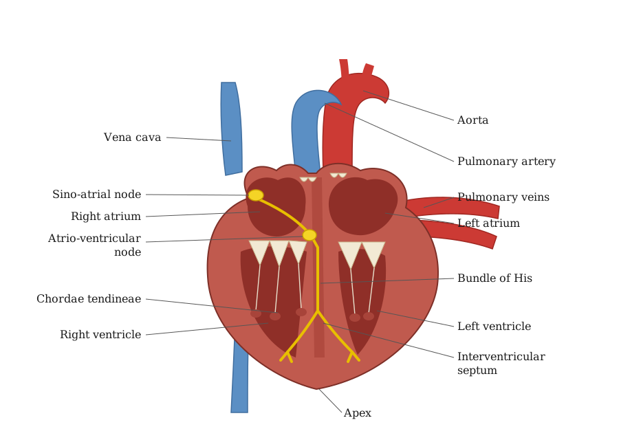
Drawn by us to help you understand the concept clearly, and verified to make sure it's accurate. For exams, practice from your textbook's own diagram.
This figure gives you a cut-away view of the human heart so that you can see how its four chambers and their partitions are arranged. Notice the two smaller upper chambers, the atria, sitting above the two larger, thicker-walled lower chambers, the ventricles. Trace the wall running down the middle: the thin inter-atrial septum separates the right atrium from the left atrium, while the much thicker inter-ventricular septum keeps the two ventricles apart. Between each atrium and the ventricle beneath it lies the atrio-ventricular septum. The figure also lets you locate the valves that force blood to travel in only one direction — the tricuspid valve between the right atrium and right ventricle, and the bicuspid (mitral) valve on the left side, along with the semilunar valves guarding the openings where the great vessels leave the heart. Reading the diagram, a student should se …