Botany · Ch 7 — Molecular Basis of Inheritance
tRNA – the Adapter Molecule
tRNA – the Adapter Molecule
The story of how the genetic code is actually read is a beautiful piece of biological logic. When Francis Crick first thought about the problem, he realised something crucial: amino acids themselves have no chemical way to recognise the triplet codons on mRNA. They are just simple molecules with no 'reading' ability. So Crick predicted that there must be an adapter molecule — something that could, on one hand, read the language of nucleic acids (the codon) and, on the other hand, carry the correct amino acid.
That adapter molecule is tRNA (transfer RNA). Interestingly, tRNA was already known before the genetic code was cracked — it was originally called sRNA (soluble RNA) because it was a small, soluble form of RNA found in the cell. But its role as the adapter was assigned only later.
Structure and function of tRNA
Every tRNA molecule has two critical functional ends:
- The anticodon loop: This contains a triplet of bases that are complementary to the codon on mRNA. This is how tRNA 'reads' the code — by base-pairing with the codon.
- The amino acid acceptor end: This is the 3' end of the tRNA, where a specific amino acid gets attached. The enzyme that does this is called aminoacyl-tRNA synthetase, and the process is called charging or aminoacylation of tRNA.
tRNAs are specific for each amino acid. That is, for every one of the 20 amino acids, there is at least one dedicated tRNA that carries only that amino acid. There are no tRNAs for stop codons — termination codons are recognised by release factors, not by tRNAs.
For the start of translation, there is a special initiator tRNA that recognises the start codon AUG.
The shape of tRNA
In its secondary structure, tRNA folds into a shape that looks like a clover-leaf (this is the classic diagram you see in textbooks). But in its actual three-dimensional structure, tRNA is a compact molecule that looks like an inverted L. The anticodon loop is at one end of the L, and the amino acid acceptor end is at the other end — this arrangement brings the two functional sites into the correct positions for the ribosome to work.
Charging of tRNA — why energy is needed
Translation is the polymerisation of amino acids into a polypeptide chain. The order of amino acids is dictated by the sequence of bases in mRNA. Amino acids are joined by peptide bonds, and forming a peptide bond requires energy. So, before translation can even begin, amino acids must be activated in the presence of ATP and linked to their specific tRNA. This is the charging step. Once two charged tRNAs are brought close together on the ribosome, the formation of a peptide bond between their amino acids becomes energetically favourable. The ribosome itself acts as a catalyst — in bacteria, the 23S rRNA acts as a ribozyme (an RNA enzyme) that catalyses peptide bond formation.
The ribosome and the translation process
The ribosome is the cellular factory for protein synthesis. It consists of structural RNAs and about 80 different proteins. In its inactive state, it exists as two subunits: a large subunit and a small subunit. The large subunit has two sites where tRNAs bind — these sites allow successive amino acids to come close enough for peptide bond formation.
Translation begins when the small subunit encounters an mRNA. The translational unit on the mRNA is the sequence flanked by a start codon (AUG) and a stop codon. But an mRNA also has untranslated regions (UTRs) — sequences at both the 5' end (before the start codon) and the 3' end (after the stop codon) that are not translated but are required for efficient translation.
The process has three main phases: …
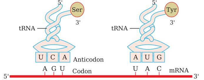
Drawn by us to help you understand the concept clearly, and verified to make sure it's accurate. For exams, practice from your textbook's own diagram.
The figure shows two separate tRNA molecules, each drawn in the classic clover-leaf secondary-structure shape — one carrying the amino acid serine (Ser) and the other carrying tyrosine (Tyr) — illustrating that each tRNA is charged with one specific amino acid at its 3′ acceptor end (shown as a small circle holding the amino-acid label).
At the bottom loop of each tRNA, three unpaired bases form the anticodon — boxed as UCA on the serine-carrying tRNA and AUG on the tyrosine-carrying tRNA. Directly below each tRNA, a single horizontal strand represents the mRNA (labelled 5′ at the left end and 3′ at the right end), carrying the matching codon in a box of its own — AGU opposite the UCA anticodon, and UAC opposite the AUG anticodon. The figure connects each anticodon triplet to its codon, showing that the tRNA reads the mRNA through complementary, antiparallel base pairing between anticodon and codon.
The figure itself keeps to these two labelled tRNA-mRNA pairs — it does not draw out or name individual internal loops of the clover-leaf beyond the anticodon loop and the acceptor end. …