Biology · Ch 17 — Locomotion and Movement
Structure of Contractile Proteins
Structure of Contractile Proteins
The banding pattern of muscle makes sense once the two contractile proteins — actin and myosin — are examined in detail. Each is not a simple rod but a built-up structure assembled from smaller units, and the way they are put together explains how a muscle is switched on and off.
Actin — the thin filament
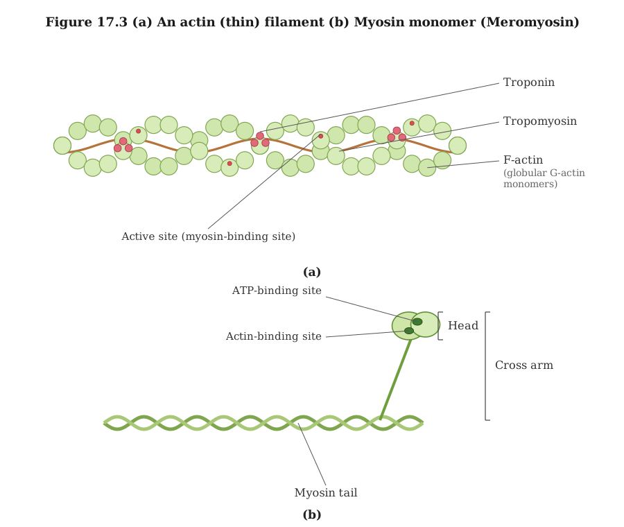
The actin, or thin, filament is a composite of several proteins:
- It is made of two strands of 'F' (filamentous) actin wound helically around each other.
- Each F-actin is itself a polymer — a chain built from many identical monomer units called 'G' (globular) actin.
- Lying alongside the F-actin strands, running the whole length of the filament, are two filaments of another protein, tropomyosin.
- At regular intervals along the tropomyosin sits a complex protein called troponin.
The arrangement matters for control: in the resting state, a subunit of troponin covers (masks) the active binding sites on actin where myosin would otherwise attach. So long as these sites are masked, myosin cannot bind and the muscle stays relaxed.
Myosin — the thick filament
The myosin, or thick, filament is also a polymerised protein, built up from many monomer units:
- The monomers that make up one thick filament are called meromyosins.
- Each meromyosin unit is built from two clearly different parts joined together -- a bulb-shaped head sitting atop a short connecting arm, and, trailing behind it, a long slender tail.
- The head together with its short arm is the heavy meromyosin (HMM).
- The tail is the light meromyosin (LMM).
- Along a polymerised myosin filament, the HMM part — the head and short arm — projects outward from the surface at a regular distance and angle from one another. This projecting portion is called the cross arm (the structure that will form the cross bridges to actin). …
Drawn by us to help you understand the concept clearly, and verified to make sure it's accurate. For exams, practice from your NCERT textbook's own diagram.
This figure shows the two filament types that slide against each other during contraction. Panel (a): the thin filament is built from two strands of F-actin twisted into a helix, and each F-actin strand is a chain of small globular G-actin units. Winding along the actin are two more proteins: tropomyosin, a long strand, and troponin, spaced at intervals. In a resting muscle these cover the active sites on actin, the spots where myosin would attach. Panel (b): a thick filament is built by stacking together many identical monomer units called meromyosins, and this panel shows just one such monomer. It has a long helical tail and, at one end, a short arm topped by a globular head -- the head-plus-arm portion together is called the cross arm. The head is the working end: it carries a binding site for ATP and a separate site for actin, and once …