Q.Differentiate between globular and fibrous proteins.
🔒You're viewing a preview — the full solution, concept, methods & PYQ mapping are locked.
🔒 Start your 14-day free trial to unlock the full solution →Concept understanding — Protein Structure Levels
Protein Structure Levels: From a String to a Working Machine
Imagine you have a long string of beads. Each bead is a different colour, and the order of colours is fixed. If you just lay that string on a table, it's a floppy, useless line. But if you could somehow make that string fold itself into a tiny, precise 3D shape — say, a key that fits a specific lock — you'd have something that actually does a job. That's exactly what a protein is.
A protein starts as a long chain of smaller units called amino acids. There are 20 different kinds, each with a unique side chain (the "colour" of the bead). The exact sequence of these amino acids is determined by your DNA. But a protein isn't just a chain — it's a chain that folds into a specific shape, and that shape determines what the protein does. If the shape is wrong, the protein can't work.
The folding happens in stages, and we call these stages the four levels of protein structure.
Level 1: Primary Structure — The Sequence
This is the simplest level: just the linear order of amino acids in the chain, linked by peptide bonds. Think of it as the sentence written in the language of proteins.
Primary structure = the sequence of amino acids from the N-terminus (start) to the C-terminus (end).
Why does this matter? Because the sequence determines everything else. Change one amino acid in a critical spot, and the entire protein can misfold. Example: sickle cell anaemia is caused by a single amino acid swap in haemoglobin — valine replaces glutamic acid at position 6. One bead out of hundreds changes colour, and the whole protein folds wrong.
Level 2: Secondary Structure — Local Folding Patterns
The chain doesn't stay straight. Hydrogen bonds form between the backbone atoms (not the side chains) of nearby amino acids. These bonds cause the chain to twist or fold into regular, repeating patterns.
Two common patterns:
- Alpha helix (α-helix): The chain coils like a spring or a spiral staircase. Hydrogen bonds form between every 4th amino acid, holding the coil tight.
- Beta sheet (β-sheet): The chain folds back and forth like a pleated fan. Hydrogen bonds form between adjacent segments, creating a flat, sheet-like structure.
Secondary structure is stabilised entirely by hydrogen bonds between the carbonyl oxygen of one amino acid and the amide hydrogen of another — both part of the peptide backbone. Side chains stick out and don't participate.
These patterns are local — they happen in short stretches of the chain. A single protein can have multiple α-helices and β-sheets separated by loops.
Level 3: Tertiary Structure — The Global 3D Shape
Now the whole chain folds into its final, compact, three-dimensional shape. This is where the protein becomes functional. The tertiary structure is stabilised by interactions between the side chains of amino acids that may be far apart in the sequence but come close in space.
What holds it together?
- Hydrophobic interactions: Nonpolar side chains cluster together in the protein's interior, away from water.
- Hydrogen bonds: Between polar side chains.
- Ionic bonds: Between positively and negatively charged side chains.
- Disulfide bridges: Covalent bonds between the sulfur atoms of two cysteine amino acids — these are strong and lock parts of the chain together.
- Van der Waals forces: Weak attractions between closely packed atoms.
A common mistake: thinking tertiary structure is just "more secondary structure." It's not. Secondary structure is local folding; tertiary structure is the global arrangement of the entire chain, including how helices and sheets pack together.
Level 4: Quaternary Structure — Multiple Chains Working Together
Some proteins are made of more than one polypeptide chain. Each chain is a separate subunit, and the quaternary structure describes how these subunits assemble into a functional complex. …
Why this formula?
Protein Structure Levels: Understanding the "Why" Behind the Hierarchy
Protein structure is not defined by a single formula, but by a logical hierarchy of organization. Each level builds on the previous one, and the "formulas" here are really principles of molecular interaction that explain why proteins fold the way they do.
Let's break down each level and the reasoning behind its key features.
1. Primary Structure: The Sequence "Formula"
What it is: The linear sequence of amino acids linked by peptide bonds.
Key "formula":
Protein=NH2-[Amino Acid]1-[AA]2-...-[AA]n-COOH
Why this holds:
- Peptide bond formation is a condensation reaction:
-COOH+NH2-→-CO-NH-+H2O
- This bond is rigid and planar due to resonance (partial double-bond character). This restricts rotation, which directly influences higher-order folding.
- The sequence is determined by DNA (genetic code). Every change in sequence can alter the entire structure — this is why a single mutation (e.g., sickle cell anemia: Glu → Val at position 6) can cause disease.
Exam insight: The primary structure is the only level that is covalently determined. All higher levels are non-covalent interactions.
2. Secondary Structure: Local Folding Patterns
Key patterns: α-helix and β-pleated sheet.
Why these form — the hydrogen bond "formula":
The α-helix
- Hydrogen bonds form between the carbonyl oxygen (C=O) of residue n and the amide hydrogen (N-H) of residue n+4.
- Why n+4? This spacing allows the backbone to coil into a right-handed helix with exactly 3.6 amino acids per turn.
- Reasoning: The peptide bond's planar nature forces the backbone into a specific geometry. The n+4 pattern maximizes H-bonding while minimizing steric clashes.
The β-sheet
- Hydrogen bonds form between adjacent strands (either parallel or antiparallel).
- Why not n+4? The backbone is extended (pleated), so H-bonds occur between different segments, not within the same chain.
Key formula (Ramachandran plot):
Only certain backbone dihedral angles (ϕ,ψ) are allowed:
- α-helix: ϕ≈−57∘, ψ≈−47∘
- β-sheet: ϕ≈−130∘, ψ≈+130∘
Why these angles? Steric hindrance — atoms cannot overlap. The Ramachandran plot shows the only regions where no two atoms clash.
3. Tertiary Structure: The 3D Fold
Key "formula": The hydrophobic effect drives folding.
Why this holds:
- Water molecules form a cage-like structure around nonpolar (hydrophobic) side chains. This is entropically unfavorable (water loses freedom).
- To minimize this, hydrophobic side chains cluster together in the protein's core, away from water.
- Result: The protein collapses into a compact globule, with polar/charged residues on the surface.
Supporting interactions (the "glue"):
| Interaction | Why it matters |
|---|---|
| Hydrogen bonds | Between side chains (e.g., Ser–Glu) |
| Ionic bonds | Between charged groups (e.g., Lys–Asp) |
| Van der Waals forces | Close packing of atoms |
| Disulfide bridges | Covalent S–S bonds (only in oxidizing environments) |
Why not just one formula? Tertiary structure is unique to each protein — it's the sum of all these interactions, not a single equation.
4. Quaternary Structure: Multiple Subunits
Key "formula":
Functional protein=∑i=1nSubuniti
Why this holds:
- Some proteins need multiple polypeptide chains to function (e.g., hemoglobin: α2β2). …
Concept: Protein Structure Levels — the distinction arises from the overall shape and function determined by the folding of polypeptide chains.
Reasoning:
- Globular proteins fold into compact, spherical shapes due to hydrophobic side chains burying inward and hydrophilic ones facing outward. This makes them water-soluble and suited for dynamic roles like enzymes, hormones, and transporters.
- Fibrous proteins form long, rod-like or sheet-like structures, often with repetitive amino acid sequences. They are insoluble in water and serve structural or protective functions (e.g., collagen, keratin, elastin). …
Globular proteins are compact, roughly spherical, and water-soluble, with tertiary structure dominated by hydrophobic cores and polar surfaces. Fibrous proteins are elongated, rod-like, and water-insoluble, with secondary structure (often α-helix or β-sheet) forming long fibres. The key difference lies in shape, solubility, and function — globular proteins are dynamic (enzymes, carriers), fibrous proteins are structural (collagen, keratin).
Why This Distinction Matters
Proteins are not just random chains of amino acids — their three-dimensional shape dictates what they can do. The difference between globular and fibrous proteins is one of the most fundamental classifications in biochemistry, and it comes down to how the polypeptide chain folds.
Think of it this way: a globular protein is like a crumpled ball of yarn — compact, with a defined inside and outside. A fibrous protein is like a stretched rope — long, repetitive, and designed to bear tension.
The Core Concepts
Globular proteins fold into compact, roughly spherical shapes. Their hydrophobic (water-fearing) amino acids tuck inside, away from water, while hydrophilic (water-loving) ones stay on the surface. This makes them soluble in water and able to move freely in cells. They are the workhorses — enzymes, hormones, transporters.
Fibrous proteins are elongated and often form long fibres or sheets. They are built from repetitive sequences that favour regular secondary structures (like the α-helix in keratin or the β-sheet in silk fibroin). They are insoluble in water and provide mechanical strength — collagen in tendons, elastin in skin, keratin in hair.
A common mistake is to think that all proteins with α-helices are fibrous. Haemoglobin is full of α-helices but is globular — it's the overall shape and solubility, not just secondary structure, that defines the class.
Step-by-Step Comparison
1. Shape and Overall Structure
- Globular: The polypeptide chain folds into a compact, roughly spherical shape. Multiple types of secondary structure (α-helices, β-sheets, loops) pack together. The tertiary structure is complex and often includes a hydrophobic core.
- Fibrous: The chain is extended, often forming long rods or sheets. Secondary structure is usually uniform — either all α-helix (keratin, myosin) or all β-sheet (silk fibroin). Quaternary structure involves many chains twisting together into cables.
2. Solubility in Water
- Globular: Soluble. Polar and charged side chains on the surface interact with water. The hydrophobic core is buried.
- Fibrous: Insoluble. The surface is dominated by hydrophobic side chains, and the repetitive sequences often lack polar residues. They are designed to stay put, not float around.
If you see a protein that dissolves in water and forms a clear solution, it's almost certainly globular. If it's tough, stringy, and won't dissolve, it's fibrous.
3. Amino Acid Composition
- Globular: A balanced mix of polar, charged, and hydrophobic amino acids. The sequence is irregular, allowing complex folding.
- Fibrous: Highly repetitive sequences. For example, collagen has the repeating tripeptide Gly-X-Y (where X is often proline, Y is often hydroxyproline). Keratin has a heptad repeat that favours coiled-coil formation.
4. Function …
Method: Structural & Functional Comparison Table
This method uses a side-by-side contrast based on shape, solubility, function, and structural level — the four pillars the exam expects.
Steps
1. Identify the shape (tertiary/quaternary structure)
- Globular: folded into a compact, spherical shape.
- Fibrous: elongated, thread-like, often forming sheets or fibers.
2. Compare solubility in water
- Globular: soluble — polar side chains face outward.
- Fibrous: insoluble — hydrophobic side chains dominate.
3. Note the primary function
- Globular: metabolic roles — enzymes, hormones, transporters.
- Fibrous: structural roles — support, elasticity, strength.
4. Check the level of protein structure involved
- Globular: often tertiary (single chain) or quaternary (multiple subunits).
- Fibrous: typically secondary structure (e.g., α-helix, β-sheet) repeated over long stretches.
Final Comparison Table
| Feature | Globular | Fibrous |
|---------|----------|---------| …
Here is a breakdown of the common mistakes students make when differentiating between globular and fibrous proteins, and how to avoid them for your exams.
🧠 The Core Concept: Why This Distinction Matters
Before we list mistakes, remember the why. The difference isn't just a list of facts; it's about function following form.
- Globular Proteins: Think "spheres" or "balls." They are folded into a compact, roughly spherical shape. This shape is crucial because it creates pockets and active sites for binding other molecules (e.g., enzymes, hormones, antibodies). They are usually water-soluble and mobile.
- Fibrous Proteins: Think "rods" or "sheets." They are long, thread-like, and often have a repeating secondary structure (like alpha-helices or beta-sheets). This shape provides structural support, strength, and elasticity. They are usually water-insoluble and static.
✗ Common Mistake #1: Confusing Solubility with the Reason for Solubility
The Mistake: Students simply state "globular proteins are soluble" and "fibrous proteins are insoluble" without explaining why.
Why it's Wrong: The exam often tests the reason. The solubility is a direct consequence of their structure.
How to Avoid It: Always link solubility to the position of hydrophobic and hydrophilic amino acids.
- Globular: Hydrophobic (water-fearing) amino acids are folded inside the core, away from water. Hydrophilic (water-loving) amino acids are on the surface, interacting with water. This makes them soluble.
- Fibrous: They have a high proportion of hydrophobic amino acids on their surface as well, making them insoluble in water.
Exam-Ready Phrase: "Globular proteins are water-soluble because their hydrophobic R-groups are buried in the interior, while hydrophilic R-groups are on the surface."
✗ Common Mistake #2: Forgetting the "Function Follows Form" Link
The Mistake: Listing functions (e.g., "enzymes are globular") without explaining why that shape is necessary for that function.
Why it's Wrong: The exam wants you to connect structure to function.
How to Avoid It: For each function, ask: "Why does this function require a spherical shape or a fibrous shape?"
- Globular (e.g., Enzyme): The spherical shape creates a specific active site (a pocket) that binds a specific substrate. The 3D folding is critical for catalysis.
- Fibrous (e.g., Collagen): The long, rope-like structure provides tensile strength (resistance to pulling). The repeating structure allows for tight packing and cross-linking.
Exam-Ready Phrase: "The compact, spherical shape of globular proteins allows for the formation of specific binding sites (e.g., active sites in enzymes), whereas the elongated, rod-like shape of fibrous proteins provides structural support and mechanical strength."
✗ Common Mistake #3: Mixing Up Examples
The Mistake: Listing an example in the wrong category (e.g., saying "collagen is a globular protein").
Why it's Wrong: This is a direct loss of marks.
How to Avoid It: Memorize the classic examples for each category. Use a simple mnemonic or association.
| Category | Key Examples | Mnemonic / Association |
|---|---|---|
| Globular | Enzymes (e.g., pepsin, trypsin), Hemoglobin, Myoglobin, Immunoglobulins (antibodies), Insulin, Albumin | Globular = Go Everywhere (mobile, soluble). Think of Hemoglobin carrying oxygen. |
| Fibrous | Collagen (skin, bones, tendons), Keratin (hair, nails, skin), Elastin (blood vessels, lungs), Fibrin (blood clotting), Myosin (muscle) | Fibrous = Firm Structure. Think of Collagen in Connective tissue. |
✗ Common Mistake #4: Ignoring the Role of Secondary Structure …
Showing the 12 most recent of 17 on this concept.
- AP EAPCET 2026Set ap-2026-05-19-FN1 markMCQQ.Choose the correct statements A) The first amino acid in a protein chain is in the left end and termed as C-terminal amino acids. B) Long protein chain folded upon itself like a hollow woolen ball is called tertiary structure. C) In general, only right handed helices are observed in protein. D) In general, polysaccharides will have glycosidic bond formed by dehydration. (A) A, B and C (B) B, C and D (C) A, C and D (D) A, B and D
›Reveal solutionSolution
Of four claims about protein/polysaccharide structure, only the "first amino acid = C-terminal" claim is wrong (it should be N-terminal); tertiary structure, right-handed helices, and dehydration-formed glycosidic bonds are all correct.
Concept and Intuition
A polypeptide has directionality: one end carries a free –NH₂ group (the N-terminus, conventionally drawn/numbered first, "left" end) and the other a free –COOH group (the C-terminus, "right" end). Beyond primary structure (sequence) and secondary structure (regular local folding like the α-helix, predominantly right-handed in natural proteins), further compact 3-D folding into a globular shape is tertiary structure. Separately, polysaccharides are built by joining monosaccharide units via glycosidic bonds, formed through dehydration (loss of a water molecule per bond), just like peptide bonds in proteins.
Step-by-Step Solution
- A: the residue with the free amino group is by convention the FIRST/N-terminal amino acid (drawn on the left) — calling it "C-terminal" is a direct mix-up of the two termini. FALSE.
- B: further folding of an already-folded (secondary structure) polypeptide chain into a compact, roughly spherical, hollow-ball-like shape is precisely the description used for tertiary structure. TRUE. …
- AP EAPCET 2025Set eng-2025-05-23-FN1 markMCQQ.Consider the following. Statement-I : Primary structure of protein represents its constitution Statement-II : α-Helix and β-pleated sheet structure of protein represent tertiary structure of it Correct answer is (A) Both statement-I and statement-II are correct (B) Both statement-I and statement-II are not correct (C) Statement-I is correct, but statement-II is not correct (D) Statement-I is not correct, but statement-II is correct
›Reveal solutionSolution
This tests the protein structure hierarchy: primary = sequence (constitution), secondary = local folding patterns like α-helix/β-sheet, tertiary = overall 3-D folding. Statement-I is right; Statement-II wrongly calls a secondary-structure feature 'tertiary'.
Concept and Intuition
Proteins are described at four structural levels: primary (linear amino-acid sequence — the actual covalent 'constitution' of the molecule), secondary (regular, repeating local folding patterns held by hydrogen bonds along the peptide backbone — the α-helix and β-pleated sheet are the two classic examples), tertiary (the overall three-dimensional folding of the whole chain, stabilised by disulfide bonds, hydrogen bonds, and hydrophobic interactions among side chains), and quaternary (arrangement of multiple polypeptide subunits). Recognising which level a named feature belongs to is the entire test here.
Step-by-Step Solution
- Statement-I: the primary structure is defined as the sequence in which amino acids are linked via peptide bonds — this is indeed the molecule's basic constitution (which amino acid, in which order). True. …
- AP EAPCET 2025Set ap-2025-05-19-AN1 markMCQQ.During the denaturation of proteins, which of the following structures will remain intact? (A) Secondary & tertiary (2∘, 3∘) (B) Tertiary only (3∘) (C) Primary only (1∘) (D) Primary and secondary (1∘, 2∘)
›Reveal solutionSolution
Denaturation breaks the weak interactions maintaining secondary/tertiary folding, but the covalent peptide-bond backbone (primary structure) survives untouched.
Concept and Intuition
Protein structure is organised in levels: primary (amino-acid sequence, held by strong covalent peptide bonds), secondary (α-helices/β-sheets, held by hydrogen bonds), and tertiary (overall 3-D fold, held by a mix of hydrogen bonds, hydrophobic effects, ionic interactions, and disulfide bridges). Denaturing agents (heat, extreme pH, chaotropic agents) disrupt these weak non-covalent interactions but do not have enough energy to break the strong covalent peptide bonds of the backbone.
Step-by-Step Solution
- Denaturation unfolds the protein by breaking the hydrogen bonds/hydrophobic interactions responsible for secondary structure.
- It also destroys tertiary structure (loss of the specific 3-D globular shape), since tertiary structure depends on similarly weak interactions (plus occasionally disulfide bonds, which some strong denaturants can also break). …
- AP EAPCET 2025Set ap-2025-05-19-FN1 markMCQQ.Match the following List-I A) Glycosidic bond B) Hollow woollen ball folding C) Enzyme D) Esterbond List-II I) Trypsin II) Bond between phosphate and 5th carbon of sugar III) tertiary structure of protein IV) formed by dehydration (A) A-II, B-I, C-IV, D-III (B) A-IV, B-III, C-I, D-II (C) A-III, B-II, C-I, D-IV (D) A-IV, B-I, C-II, D-III
›Reveal solutionSolution
Matching each biomolecule term to its correct description gives A-IV, B-III, C-I, D-II — answer (B).
Concept and Intuition
- Glycosidic bond: forms between two monosaccharides via a dehydration (condensation) reaction, releasing a water molecule.
- The image of a protein folding "like a hollow woollen ball" is a common descriptive analogy for a protein's tertiary structure — the overall 3D folding of a single polypeptide chain.
- Trypsin is a well-known digestive protease and is the standard textbook example cited for "enzyme."
- The ester bond joining a phosphate group to the 5′-carbon hydroxyl of a sugar (as in nucleotides) is a phosphoester linkage.
Step-by-Step Solution
- A) Glycosidic bond → IV (formed by dehydration).
- B) "Hollow woollen ball" folding → III (tertiary structure of protein).
- C) Enzyme → I (Trypsin, a textbook enzyme example). …
- AP EAPCET 2025Set ap-2025-05-19-FN1 markMCQQ.Choose the correct statements among the following I. In exothermic reactions 'P' is at a lower level than 'S'. II. Enzyme activity can be affected by temperature etc. with alteration of tertiary structure of protein. III. Low temperature denature the protein. IV. Ligases can remove the groups from substrate (A) I, II (B) III, IV (C) II, IV (D) I, III
›Reveal solutionSolution
Exothermic reactions do place products at lower energy than substrates, and temperature does affect activity via tertiary-structure changes — both true. Low temperature inactivates (not denatures) enzymes, and ligases join (not remove) — both false. Correct set: I, II — option (A).
Concept and Intuition
Enzyme-catalysed reaction energetics: in an exothermic reaction, the products (P) are at a lower free-energy level than the substrates (S), with the difference released as heat — this is basic thermodynamics of catalysed reactions and is independent of the enzyme itself (the enzyme only lowers the activation energy, not the net energy change). Enzyme activity is highly temperature-sensitive because temperature affects the enzyme's three-dimensional (tertiary) structure, which determines the shape of its active site; this is why activity rises with temperature up to an optimum and then falls off as heat begins to disrupt this structure (denaturation) at high temperatures. Crucially, LOW temperature does not denature an enzyme — it merely reduces the kinetic energy of molecules, slowing the reaction reversibly (activity resumes when temperature rises again); denaturation (irreversible loss of structure) is a HIGH-temperature (or extreme pH) phenomenon. Ligases are enzymes that join two molecules together (often using ATP, e.g., DNA ligase sealing nicks) — they do not remove groups from a substrate; that role belongs to hydrolases/lyases.
Step-by-Step Solution
- Statement I: In exothermic reactions, product (P) is at a lower energy level than substrate (S) — TRUE, by definition of an exothermic process (energy released, product lower in energy). …
- AP EAPCET 2024Set eng-2024-05-21-FN1 markMCQQ.Which of the following structure of proteins represents its constitution? (A) Secondary structure (B) Quaternary structure (C) Primary structure (D) Tertiary structure
›Reveal solutionSolution
A protein's constitution — the linear sequence of amino acid residues — is defined as its primary structure.
Concept and Intuition
Protein structure is described at four levels: primary (amino-acid sequence, held by peptide bonds), secondary (local folding like α-helix/β-sheet, via H-bonds), tertiary (overall 3-D fold, via various side-chain interactions), and quaternary (arrangement of multiple polypeptide subunits). "Constitution" refers to which atoms/residues are bonded to which — i.e. connectivity — which is captured entirely by the primary structure; the higher-order structures describe spatial conformation, not constitution.
Step-by-Step Solution
- Recall the four levels of protein structure and what each describes.
- "Constitution" = the sequence/connectivity of amino acid units — matches the definition of primary structure exactly. …
- AP EAPCET 2023Set ap-2023-05-22-AN1 markMCQQ.Enzymes are denaturated by (A) Low pH (B) High substrate concentration (C) Low temperature (D) heat
›Reveal solutionSolution
Enzymes lose their catalytic 3-D shape and activity when heated — heat is the classic, universally correct denaturing agent among the given options.
Concept and Intuition
An enzyme's activity depends entirely on its precise tertiary (3-D) structure, especially the shape of its active site. This structure is held together by relatively weak, non-covalent interactions (hydrogen bonds, ionic bonds, hydrophobic interactions) that are easily disrupted by excess thermal energy, causing the protein to unfold and lose its functional shape — this is denaturation.
Step-by-Step Solution
- (A) Low pH can denature enzymes only in extreme cases; mild pH shifts merely reduce activity reversibly rather than denaturing outright, so it's not the cleanest universal answer.
- (B) High substrate concentration does not denature an enzyme; at most it can cause substrate inhibition, a different, reversible phenomenon.
- (C) Low temperature slows enzyme activity (reduces molecular motion/collision frequency) but does not denature the enzyme — activity resumes on warming. …
- AP EAPCET 2023Set eng-2023-05-15-FN1 markMCQQ.The structure of Gly-Ala is (A) [FIGURE] (a dipeptide drawn with an alanine residue - H2N−CH(CH3)−CO− - joined via an amide bond to a glycine residue - −NH−CH2−CO2H; i.e. Ala-Gly) (B) [FIGURE] (a dipeptide drawn with a glycine residue - H2N−CH2−CO− - joined via an amide bond to an alanine residue - −NH−CH(CH3)−CO2H; i.e. Gly-Ala) (C) [FIGURE] (a dipeptide drawn with an alanine residue - H2N−CH(CH3)−CO− - joined via an amide bond to a serine residue - −NH−CH(CH2OH)−CO2H; i.e. Ala-Ser) (D) [FIGURE] (a dipeptide drawn with a serine residue - H2N−CH(CH2OH)−CO− - joined via an amide bond to an alanine residue - −NH−CH(CH3)−CO2H; i.e. Ser-Ala)
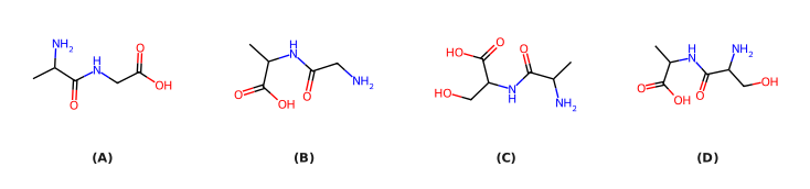
›Reveal solutionSolution
The structure of Gly-Ala is a dipeptide where glycine (no side chain) is the N‑terminal residue and alanine (methyl side chain) is the C‑terminal residue. The correct option is (B).
Concept and Intuition
Proteins and peptides are built from amino acids linked by peptide (amide) bonds. The name of a dipeptide is written from the N‑terminus (free amino group) to the C‑terminus (free carboxyl group). So “Gly-Ala” means glycine is at the left (N‑terminal) end, and alanine is at the right (C‑terminal) end.
- Glycine (Gly) has a hydrogen atom as its side chain — so its α‑carbon has no branch (just –H).
- Alanine (Ala) has a methyl group (–CH₃) as its side chain — so its α‑carbon carries a small branch.
Thus, in the correct structure:
- The leftmost part (H₂N–) is attached to a carbon with no side chain (glycine).
- That carbon is linked via an amide bond (–CO–NH–) to the α‑carbon of alanine, which does have a methyl branch.
- The rightmost part is the free carboxyl group (–CO₂H) of alanine.
Step‑by‑Step Reasoning
-
Identify the residues from the name
“Gly-Ala” tells us the sequence: Glycine first, then Alanine.
- Glycine: H₂N–CH₂–COOH (side chain = H)
- Alanine: H₂N–CH(CH₃)–COOH (side chain = CH₃)
-
Form the peptide bond
The carboxyl group of glycine reacts with the amino group of alanine, releasing water. The resulting dipeptide is:
H2N−CH2−C(=O)−NH−CH(CH3)−CO2H
Notice:
- Left side (N‑terminus): H₂N–CH₂– (glycine, no branch)
- Middle: amide bond (–CO–NH–)
- Right side (C‑terminus): –CH(CH₃)–CO₂H (alanine, methyl branch)
- Match with the given options
- Option (A): Left carbon has a methyl branch (alanine), right carbon has no branch (glycine) → Ala-Gly, not Gly-Ala.
- Option (B): Left carbon has no branch (glycine), right carbon has a methyl branch (alanine) → Gly-Ala. …
- AP EAPCET 2023Set eng-2023-05-17-AN1 markMCQQ.Which of the following are fibrous proteins? Keratin (A) Insulin (B) Myosin (C) Albumin (D) (A) A, B (B) A, C (C) B, D (D) C, D
›Reveal solutionSolution
Fibrous proteins are structural, elongated molecules; keratin and myosin are fibrous, while insulin and albumin are globular.
Concept and Intuition
Proteins are broadly classed by shape/function: fibrous proteins are long thread-like molecules (often helices/sheets) giving mechanical strength; globular proteins fold compactly and perform dynamic biochemical roles.
Step-by-Step Solution
- Keratin (A): structural protein in hair/nails/wool - fibrous.
- Insulin (B): small globular hormone regulating blood glucose - globular.
- Myosin (C): motor protein of muscle, forms long filaments - fibrous. …
- AP EAPCET 2022Set ap-2022-07-11-FN1 markMCQQ.Hemoglobin in man is made up of (A) 4 Polypeptide chains and 4 haeme molecules (B) 2 Polypeptide chains and 2 haeme molecules (C) 4 Polypeptide chains and one haeme molecules (D) 2 Polypeptide chains and 1 haeme molecule
›Reveal solutionSolution
This tests the quaternary structure of haemoglobin; the answer is 4 polypeptide chains and 4 haeme molecules.
Concept and Intuition
Haemoglobin is a globular, tetrameric protein. Each of its 4 globin (polypeptide) subunits folds around one haem prosthetic group, whose central Fe2+ ion reversibly binds one O2 molecule. Because oxygen-binding happens per haem group and there are 4 haem groups (one per chain), a single haemoglobin molecule can carry up to 4 O2 molecules — this is also the structural basis of cooperative binding (the sigmoidal O2-dissociation curve).
Step-by-Step Solution
- Adult haemoglobin (HbA) = α2β2, i.e., 2 alpha-globin chains + 2 beta-globin chains = 4 polypeptide chains total.
- Each chain has exactly one haem group embedded in it — so 4 chains → 4 haem groups.
- This directly matches option (A): 4 polypeptide chains and 4 haeme molecules. …
- AP EAPCET 2022Set ap-2022-07-12-AN1 markMCQQ.Which among the following are peptide based hormones?(i) Estrogen(ii) Insulin(iii) Endorphin(iv) Androgen (A)(i) and(iv) (B)(ii) and(iii) (C)(i) and(iii) (D)(ii) and (iv)
›Reveal solutionSolution
Among the four, only insulin and endorphin are peptide hormones; estrogen and androgen are steroids.
Concept and Intuition
Hormones are chemically classified as steroids (derived from cholesterol, e.g., sex hormones), peptides/proteins (chains of amino acids, e.g., insulin, glucagon), or amino-acid derivatives (e.g., thyroxine, adrenaline). Knowing this classification directly answers such questions.
Step-by-Step Solution
- Estrogen (i) — a steroid hormone (female sex hormone), not peptide-based.
- Insulin (ii) — a well-known peptide hormone made of two polypeptide chains (A and B chains).
- Endorphin (iii) — an endogenous opioid peptide, hence peptide-based. …
- AP EAPCET 2022Set eng-2022-07-06-AN1 markMCQQ.Which of the following is not true about the denaturation of protein? (A) 2∘ structure get destroyed (B) 3∘ structure get destroyed (C) 1∘ structure get destroyed (D) Bioactivity is lost
›Reveal solutionSolution
Denaturation unfolds a protein by breaking the weak interactions maintaining its 2°/3° shape and destroys its biological activity, but it leaves the covalent peptide-bond backbone (the primary structure) intact.
Concept and Intuition
A protein's structure is organised in levels: primary (the amino-acid sequence, held together by strong covalent peptide bonds), secondary (α-helices/β-sheets, held by H-bonds along the backbone), and tertiary (the overall 3-D fold, held by H-bonds, hydrophobic interactions, ionic bonds, and disulfide bridges between side chains). Denaturation — by heat, extreme pH, heavy metals, or other agents — supplies enough energy/disruption to break the weak interactions holding 2°/3° structure, so the protein unfolds into a random coil and loses its specific shape. Since biological activity (e.g. enzyme catalysis) depends entirely on that specific 3-D shape, activity is lost. But the sequence of amino acids joined by covalent peptide bonds is not affected by these mild conditions — that would require actually breaking covalent bonds (hydrolysis), a chemically much more demanding process.
Step-by-Step Solution
- Recall what denaturation physically does: it disrupts H-bonds, hydrophobic interactions, and other weak forces stabilising the folded state. …
🎓Unlock everything free for 14 days
- ✓Full step-by-step solutions
- ✓Concept-first explanations
- ✓Methods, shortcuts & mistakes
- ✓PYQ mapping + timed mock tests
Full access for 14 days. No credit card required.