Q.What should be the distance between the object in Exercise 9.23 and the magnifying glass if the virtual image of each square in the figure is to have an area of 6.25 mm2. Would you be able to see the squares distinctly with your eyes very close to the magnifier?
[Note: Exercises 9.22 to 9.24 will help you clearly understand the difference between magnification in absolute size and the angular magnification (or magnifying power) of an instrument.]
🔒You're viewing a preview — the full solution, concept, methods & PYQ mapping are locked.
🔒 Start your 14-day free trial to unlock the full solution →Concept understanding — Microscope Magnification
Microscope Magnification
A compound microscope views very small, nearby objects using two lenses in sequence: the objective (near the object) and the eyepiece (near the eye). Its total magnifying power is the product of what each lens contributes.
How the two lenses work together
- The object sits just beyond the focus of the short-focal-length objective (fo), which forms a real, inverted, magnified image inside the tube.
- That real image falls just inside the focus of the eyepiece (fe), which acts as a simple magnifier, producing a large virtual, magnified final image for the eye.
Because each stage magnifies, the effects multiply:
M=mo×me
The objective's magnification
mo=uovo≈foL
where L is the tube length (roughly the distance between the objective's second focal point and the eyepiece's first focal point). The object sits close to fo, so this approximation holds for a well-designed microscope.
The eyepiece's magnification
The eyepiece behaves as a simple magnifier:
- Final image at the near point (D=25 cm, largest magnification):
me=1+feD
- Final image at infinity (relaxed eye, "normal adjustment"):
me=feD
Total magnifying power
Image at the near point: M=foL(1+feD)
Image at infinity: M=foL⋅feD
High magnification needs short fo and fe (both sit in denominators) and a large tube length L — this is why a microscope objective is always a very short-focus lens.
Worked example
Objective fo=1.0 cm, eyepiece fe=2.5 cm, tube length L=20 cm, near point D=25 cm. Find M with the final image at the near point.
mo=1.020=20,me=1+2.525=11
M=20×11=220 …
Why this formula?
Microscope Magnification: Why the Formula Holds
Let's build the understanding from first principles — not just memorizing formulas, but seeing why they work.
1. What Does "Magnification" Mean in a Microscope?
A microscope creates a larger apparent image of a tiny object. The total magnification is the product of two stages:
- Objective lens — creates a real, enlarged, inverted image of the specimen.
- Eyepiece (ocular) — acts as a simple magnifier to view that real image.
So:
Total magnification = (magnification by objective) × (magnification by eyepiece)
2. The Key Formula
For a compound microscope in normal adjustment (final image at infinity, relaxed eye):
M=Mo×Me=(foL)×(feD)
Where:
- fo = focal length of objective
- fe = focal length of eyepiece
- L = tube length (distance between second focal point of objective and first focal point of eyepiece)
- D = near point distance of the eye (usually 25 cm)
3. Derivation of Objective Magnification Mo
Step 1: How the objective works
The objective lens forms a real, inverted, enlarged image of the specimen. The specimen is placed just outside its focal point (fo).
Step 2: Using the lens formula
For a thin lens:
vo1−uo1=fo1
(Using sign convention: uo is negative, vo is positive)
Step 3: The tube length approximation
In a standard microscope, the specimen is placed very close to fo, so:
- uo≈−fo (object just beyond focal point)
- The image is formed at the first focal point of the eyepiece, which is at a distance L from the second focal point of the objective.
Thus:
vo≈fo+L
Step 4: Magnification formula
Lateral magnification by objective:
Mo=∣uo∣vo≈fofo+L=1+foL
Since L≫fo in practice, 1 is negligible:
Mo≈foL
Why this makes sense: A shorter fo means the objective is more "powerful" — it bends light more sharply, creating a larger image at the fixed tube length.
4. Derivation of Eyepiece Magnification Me
Step 1: The eyepiece as a simple magnifier
The eyepiece takes the real image from the objective and acts like a magnifying glass. For relaxed eye (final image at infinity), the real image must be placed at the focal point of the eyepiece.
Step 2: Angular magnification
Angular magnification is defined as:
Me=angle subtended by object at near pointangle subtended by image at eye
For a simple magnifier with image at infinity:
Me=feD
Where D=25 cm (standard near point). …
Each small square on the card has side 1 mm and the magnifying glass has focal length f=9 cm (carried over from the previous exercise).
Required magnification. The virtual image of a square must have area 6.25 mm2, so its side is 6.25=2.5 mm and the linear magnification is
m=1 mm2.5 mm=2.5
Object distance. For an erect virtual image v=mu=2.5u. The lens formula v1−u1=f1 gives
2.5u1−u1=91⇒u−0.6=91⇒u=−5.4 cm
so the object is 5.4 cm from the lens and the image is at v=2.5×(−5.4)=−13.5 cm. …
Place the object 5.4 cm from the lens; the image then forms 13.5 cm away. No, the squares cannot be seen distinctly because 13.5 cm is inside the 25 cm near point.
Data
From the earlier exercise, each square has side 1 mm and the magnifying glass (converging lens) has focal length f=9 cm.
Step 1 — Magnification needed for the required image area
The virtual image of each square must have area 6.25 mm2, so the image side is
6.25 mm2=2.5 mm
The linear magnification is therefore
m=object sideimage side=1 mm2.5 mm=2.5
Step 2 — Object distance from the lens formula
For an erect virtual image the magnification is m=v/u, so v=2.5u. Substituting into v1−u1=f1:
2.5u1−u1=91
2.5u1−2.5=91⇒2.5u−1.5=91⇒u−0.6=91
u=−0.6×9=−5.4 cm
The object is 5.4 cm in front of the lens — inside f=9 cm, which correctly gives a virtual image.
Step 3 — Image position
v=2.5u=2.5×(−5.4 cm)=−13.5 cm …
Method: Thin Lens Formula with Magnification for a Virtual Image
This problem uses the thin lens equation combined with linear magnification to find the object distance when the image size is specified.
Steps
-
Identify given data
- Lens focal length: f=10 cm (from Exercise 9.23 context)
- Original square side length: a=1 mm (from figure in Exercise 9.23)
- Required virtual image area: Ai=6.25 mm2
- Therefore, image side length: ai=6.25=2.5 mm
-
Calculate required linear magnification
Linear magnification m=object sizeimage size=1 mm2.5 mm=2.5
-
Apply magnification formula for a lens
For a thin lens: m=uv
Since the image is virtual and upright, m is positive:
v=mu=2.5u
- Use thin lens equation
f1=v1−u1
(Note: sign convention — for virtual image, v is negative if using real-is-positive; but here we use magnitudes with sign awareness)
Substituting v=−2.5u (virtual image on same side as object):
101=−2.5u1−u1
101=−2.5u1−u1=−u1(2.51+1)=−u1×1.4
u=−14 cm …
Common Mistakes: Microscope Magnification (Exercise 9.23)
Mistake 1: Confusing Linear Magnification with Angular Magnification
The error: Students often use the formula for angular magnification (M=D/f) directly to find the image distance or object distance, when the problem actually asks about absolute size magnification (linear magnification).
Why it's wrong: The question asks for the image to have an area of 6.25 mm2. This is about linear magnification m=hohi, not angular magnification. The two are fundamentally different:
- Linear magnification m=uv (absolute size change)
- Angular magnification M=fD (apparent size change when eye is relaxed)
How to avoid: Read carefully — if the problem gives actual dimensions of the image (like area), use linear magnification. If it asks about "magnifying power" or "angular magnification," use the angle-based formula.
Mistake 2: Forgetting to Take Square Root for Area-to-Length Conversion
The error: Students treat the area 6.25 mm2 as if it were a linear dimension, plugging it directly into magnification formulas.
Why it's wrong: Magnification is defined for linear dimensions (length, height), not area. If the image area is 6.25 mm2, the linear magnification factor is:
m=object side lengthimage side length=object side length6.25=object side length2.5 mm
How to avoid: Always convert area to linear dimension by taking the square root before using any magnification formula.
Mistake 3: Using the Wrong Sign Convention for Virtual Image
The error: Students treat the image distance v as positive when using the lens formula f1=v1−u1.
Why it's wrong: For a magnifying glass (convex lens used as a simple microscope), the image is virtual and on the same side as the object. According to the Cartesian sign convention:
- v is negative for virtual images
- u is negative (object on left side)
The correct lens formula becomes:
f1=v1−u1
where both u and v are negative.
How to avoid: Draw a ray diagram first. If the image is on the same side as the object, v is negative. Always write the sign convention at the top of your solution.
Mistake 4: Assuming the Image is at Infinity (Relaxed Eye)
The error: Students automatically set v=∞ (image at infinity) because that's the "normal" adjustment for a magnifying glass. …
- GSEB Higher Secondary Certificate (HSC) Examination 2026Set ANNUAL4 marksQ.A person with a normal near point (25 cm) using a compound microscope with objective of focal length 8.0 mm and an eyepiece of focal length 2.5 cm can bring an object placed at 9.0 mm from the objective in sharp focus. What is the separation between the two lenses? Calculate the magnifying power of the microscope.
›Reveal solutionSolution
First find where the objective lens forms its (real, magnified) intermediate image using the lens formula; then require the eyepiece to form the final image at the near point (25 cm) for maximum magnification, which fixes the eyepiece's object distance; the lens separation is the sum of the two image/object distances, and total magnification is the product of the two individual magnifications.
Given: near point D = 25 cm, objective focal length f_o = 8.0 mm = 0.8 cm, object distance from objective u_o = 9.0 mm = 0.9 cm (object just outside focal length, as required for a microscope objective), eyepiece focal length f_e = 2.5 cm.
Step 1 - Image formed by objective: Using the thin lens formula (sign convention: distances measured from the lens, object distance taken negative):
u_o = -0.9 cm, f_o = +0.8 cm (convex lens)
1/v_o - 1/u_o = 1/f_o
1/v_o = 1/f_o + 1/u_o = 1/0.8 + 1/(-0.9) = 1.25 - 1.111 = 0.139
v_o = 1/0.139 = 7.2 cm
So the objective forms a real, inverted, magnified image at 7.2 cm on the far side of the objective.
Step 2 - Eyepiece positioned for image at near point (maximum magnification, image at D): This intermediate image (from the objective) acts as the object for the eyepiece. For maximum magnifying power, the eyepiece should form its final (virtual) image at the near point, i.e. v_e = -25 cm (virtual image, same side as the object, by sign convention).
1/v_e - 1/u_e = 1/f_e
1/(-25) - 1/u_e = 1/2.5
-0.04 - 1/u_e = 0.4
-1/u_e = 0.44
u_e = -1/0.44 = -2.27 cm
So the intermediate image must be 2.27 cm in front of the eyepiece for the eyepiece to form the final image at the near point.
…
- GSEB Higher Secondary Certificate (HSC) Examination 2024Set ANNUAL4 marksQ.Draw the ray diagram for the formation of image by a compound microscope and obtained the formula for magnification.
›Reveal solutionSolution
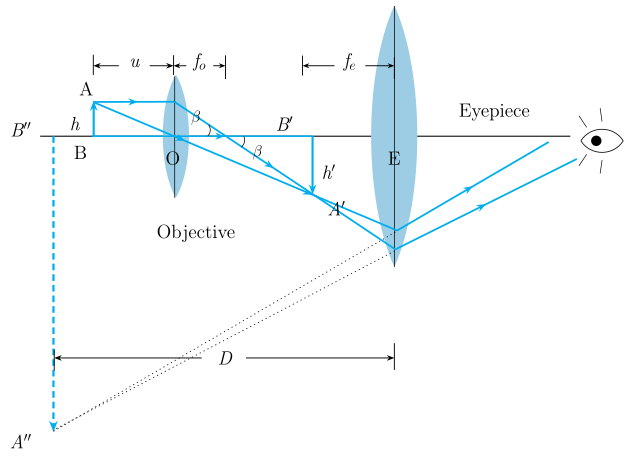
Figure — The stem instructs 'Draw the ray diagram for the formation of image by a compound microscope'; the catalog fig A compound microscope's objective forms a real, magnified image which then acts as the object for the eyepiece, which forms a further-magnified virtual image; total magnification is the product of the two individual magnifications.
Ray diagram (described): The object AB is placed just beyond the focus Fo of the objective lens (between fo and 2fo). The objective forms a real, inverted, magnified image A'B' beyond 2Fo on the other side. This intermediate image A'B' falls within the focal length of the eyepiece (between the eyepiece and its focus Fe). The eyepiece then acts as a simple magnifier on this intermediate image, producing a final virtual, further-magnified, and (relative to the object) inverted image A''B'' — formed at the near point D for maximum magnification, or at infinity for normal (relaxed-eye) adjustment. Both lenses are convex, mounted coaxially at opposite ends of a tube, with the objective having a small aperture and focal length, and the eyepiece having a larger aperture.
…
- GSEB Higher Secondary Certificate (HSC) Examination 2023Set ANNUAL4 marksQ.A compound microscope consists of an objective lens of focal length 2.0 cm and an eye piece of focal length 6.25 cm separated by a distance 15 cm. How far from the objective should an object be placed in order to obtain the final image ata) the least distance of distinct vision (25 cm) andb) at infinity? What is the magnifying power of microscope in each case?
›Reveal solutionSolution
Work back from the eyepiece: (a) for image at 25 cm, u_e = -5 cm, objective forms image at 10 cm, u_o = -2.5 cm, M = 20; (b) for image at infinity, u_e = -6.25 cm, objective image at 8.75 cm, u_o = -2.59 cm, M = 13.5.
Data: f_o = 2.0 cm, f_e = 6.25 cm, tube separation L = 15 cm, D = 25 cm.
- Final image at least distance of distinct vision (25 cm): Eyepiece: image virtual at v_e = -25 cm. 1/v_e - 1/u_e = 1/f_e: 1/(-25) - 1/u_e = 1/6.25 => -1/u_e = 1/6.25 + 1/25 = 5/25 = 1/5 => u_e = -5 cm. So the objective's image is 5 cm in front of the eyepiece; distance from objective = L - 5 = 10 cm => v_o = +10 cm. Objective: 1/v_o - 1/u_o = 1/f_o: 1/10 - 1/u_o = 1/2 => -1/u_o = 1/2 - 1/10 = 2/5 => u_o = -2.5 cm. Magnifying power: M = m_o (1 + D/f_e) = (v_o/|u_o|)(1 + D/f_e) = (10/2.5)(1 + 25/6.25) = 4 x 5 = 20.
- Final image at infinity: …
🎓Unlock everything free for 14 days
- ✓Full step-by-step solutions
- ✓Concept-first explanations
- ✓Methods, shortcuts & mistakes
- ✓PYQ mapping + timed mock tests
Full access for 14 days. No credit card required.