Q.Briefly describe the structure of human eye with the help of a labelled diagram. OR Describe the structure of human neuron with the help of a labelled diagram.
You're viewing a preview — the full solution, concept, methods & PYQ mapping are locked.
Start your 14-day free trial to unlock the full solution →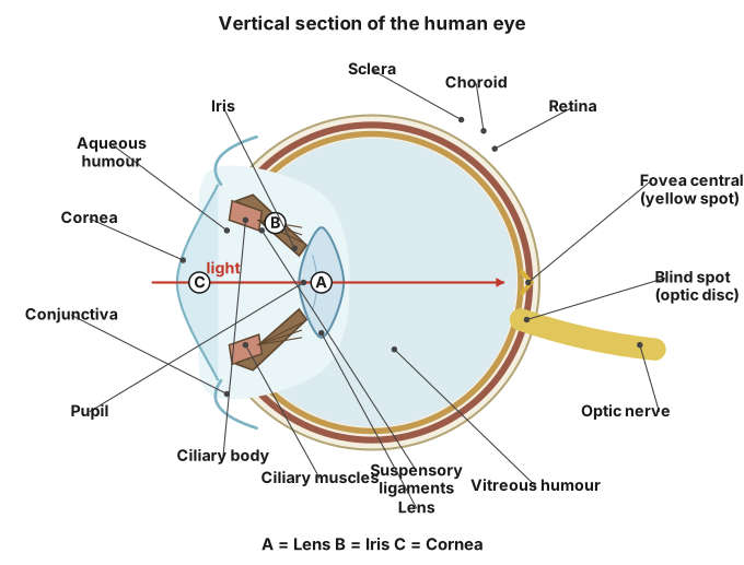
The human eye is a spherical camera-like organ built of three concentric layers (sclera-cornea, choroid-iris-ciliary body, retina) plus a lens and two humours that together focus light onto the retina, whose photoreceptors send signals to the brain via the optic nerve.
Structure of the Human Eye
The eyeball is nearly spherical and set in a bony socket (orbit) of the skull. It is made of three layers:
- Outer layer (fibrous coat): the tough, white, opaque sclera covers most of the eyeball and maintains its shape; at the front it becomes the transparent, curved cornea, through which light enters.
- Middle layer (vascular coat / uvea): the pigmented, blood-vessel-rich choroid lines most of the inside of the sclera and nourishes the retina; anteriorly it forms the ciliary body (which holds the lens in place via suspensory ligaments and controls its curvature) and the iris, the coloured, muscular diaphragm with a central opening, the pupil, that regulates the amount of light entering the eye.
- Inner layer: the retina, containing the light-sensitive photoreceptor cells — rod cells (dim-light, black-and-white vision) and cone cells (bright-light, colour vision). The point where the optic nerve leaves the retina, the blind spot, has no photoreceptors; the fovea/yellow spot, rich in cones, is the region of sharpest vision.
Inside the eyeball, behind the pupil, lies the transparent, biconvex lens, held by ciliary (suspensory) ligaments; it fine-focuses light onto the retina by changing its curvature (accommodation). The space between cornea and lens is filled with a watery fluid, the aqueous humour, and the larger chamber between lens and retina is filled with a jelly-like fluid, the vitreous humour — both help maintain the eyeball's shape and refract light.
…
Unlock everything free for 14 days
- Full step-by-step solutions
- Concept-first explanations
- Methods, shortcuts & mistakes
- PYQ mapping + timed mock tests
Full access for 14 days. No credit card required.