Q.Collect 5 examples of palindromic DNA sequences by consulting your teacher. Better try to create a palindromic sequence by following base-pair rules.
🔒You're viewing a preview — the full solution, concept, methods & PYQ mapping are locked.
🔒 Start your 14-day free trial to unlock the full solution →Concept understanding — Palindromic DNA Sequences
Imagine you are looking in a mirror. Your right hand becomes the reflection’s left hand, and your left becomes its right. The word “MALAYALAM” reads the same forwards and backwards. That mirror-like symmetry is the core idea behind a palindromic DNA sequence.
In everyday language, a palindrome is a word, phrase, or number that reads identically in both directions. DNA is a long, double-stranded molecule — think of it as a twisted ladder. Each rung of the ladder is made of two chemical letters (called bases) that pair up in a very specific way: A always pairs with T, and C always pairs with G.
Now, a palindromic DNA sequence is a stretch of DNA where the sequence of letters on one strand reads exactly the same as the sequence on the opposite strand, but in the opposite direction. Because the two strands run in opposite directions (biologists call this “antiparallel”), the palindrome is not just a simple mirror of letters — it is a mirror of the pairing.
In textbooks, you will often see a palindromic sequence written like this:
5' – GAATTC – 3'
3' – CTTAAG – 5'
Notice that if you read the top strand left to right (GAATTC) and then read the bottom strand right to left (also GAATTC), you get the same sequence. That is the palindrome.
Why does this matter? Because nature uses these sequences as recognition sites. Special proteins — especially restriction enzymes — are designed to find these exact palindromic stretches and cut the DNA at that precise spot. This is the foundation of genetic engineering:
- Restriction enzymes act like molecular scissors. They only cut at their specific palindromic sequence.
- Because the sequence is the same on both strands, the cut produces either “blunt” ends or “sticky” ends (short, single-stranded overhangs). Sticky ends are particularly useful because they can easily join with another piece of DNA that has the complementary sticky end — like two puzzle pieces.
- This allows scientists to cut DNA from one organism and paste it into the DNA of another, creating recombinant DNA.
The NCERT textbook (Class 12 Biology, Chapter 11) explicitly states: “Restriction enzymes cut the strand of DNA a little away from the centre of the palindromic site, but between the same two bases on the opposite strands.” This means the cut is not perfectly in the middle — it is offset, which creates the sticky ends. …
Palindromic DNA sequences are those that read the same on both strands when you read one strand in the 5' to 3' direction and the other strand also in the 5' to 3' direction. This happens because the sequence on one strand is identical to its complementary strand when both are read in the same direction. The key is that the base-pairing rules (A with T, and G with C) must hold, so the sequence on one strand determines the sequence on the other.
To create your own palindromic sequence, start by writing a short stretch of DNA on one strand, say 5' to 3'. Then, write its complementary strand in the 3' to 5' direction. Now, reverse the complementary strand so it reads 5' to 3'. If this reversed complementary strand matches your original strand exactly, you have a palindrome. For example, if you start with 5' GAATTC 3', its complement is 3' CTTAAG 5'. Reversing that complement gives 5' GAATTC 3' — a perfect mat …
A palindromic DNA sequence reads the same on both strands when read in the 5' to 3' direction, and it is created by following the base-pairing rules (A with T, G with C) so that the sequence on one strand is the reverse complement of the other.
Let’s begin with the idea. In everyday language, a palindrome is a word or phrase that reads the same forwards and backwards — like “MADAM” or “RACECAR.” DNA, being a double-stranded molecule, has its own version of this. A palindromic DNA sequence is one where the sequence of bases on one strand, when read from the 5' end to the 3' end, is exactly identical to the sequence on the complementary strand when it is also read from its 5' end to its 3' end. This is not just a neat trick of nature; it is a structurally important feature, especially in the context of restriction enzymes — the molecular scissors used in genetic engineering. Many restriction enzymes recognise and cut at specific palindromic sequences, which is why this concept is central to your NCERT syllabus.
To understand how to create one, you must first recall the base-pairing rules: Adenine (A) always pairs with Thymine (T), and Guanine (G) always pairs with Cytosine (C). Now, imagine you write a short sequence on one strand, say 5' – GAATTC – 3'. To get the complementary strand, you would write the partner bases: C for G, T for A, A for T, T for A, G for C, and C for G. That gives you 3' – CTTAAG – 5'. But here is the crucial step: to check for a palindrome, you must read that complementary strand in the 5' to 3' direction. So you flip it: 5' – GAATTC – 3'. It is exactly the same as the original strand. That is a palindrome.
The key is that the two strands are reverse complements of each other. If you take one strand, reverse its sequence, and then take the complement of that reversed sequence, you get back the other strand. This is what makes the sequence palindromic. …
Alternative Approach: A Faster "Outside-In Pairing" Shortcut
Writing out the full complementary strand and then reversing it (as in the main answer)
always works, but there is a quicker mental shortcut for both checking and constructing
palindromes.
Step 1: Understand the shortcut rule.
For a sequence to be palindromic, the base at position 1 (from the 5' end) must be the
Watson-Crick complement of the base at the LAST position; the base at position 2 must be
the complement of the second-last position; and so on, working inward from both ends.
Step 2: Apply it to check an existing sequence, e.g. GAATTC.
Position 1 (G) vs position 6 (C): G pairs with C -- match.
Position 2 (A) vs position 5 (T): A pairs with T -- match.
Position 3 (A) vs position 4 (T): A pairs with T -- match.
All three outside-in pairs check out, so GAATTC is palindromic -- confirmed without
writing out a second strand at all.
Step 3: Apply it to construct a new one.
Pick any base for position 1, say C; position 6 must then be G. Pick any base for …
- KCET 2025Set C-41 markMCQQ.Choose the correct sequence of steps involved in decomposition (A) Fragmentation → Leaching → Catabolism → Mineralisation → Humification (B) Fragmentation → Mineralisation → Humification → Leaching → Catabolism (C) Fragmentation → Leaching → Catabolism → Humification → Mineralisation (D) Fragmentation → Catabolism → Leaching → Humification → Mineralisation
›Reveal solutionSolution
Detritivores break the litter up, water washes out the solubles, enzymes digest the rest, the leftover resistant material becomes humus, and only then is that humus mineralised to inorganic nutrients — so mineralisation is the final step.
Step 1 — The five processes, in the order they must happen.
- Fragmentation. Detritivores (e.g. earthworms) physically break the detritus into smaller particles. This must be first: it hugely increases the surface area on which everything else acts.
- Leaching. Water percolating through the fragmented litter washes water-soluble inorganic nutrients down into the soil, where they get precipitated as unavailable salts.
- Catabolism. Bacterial and fungal enzymes degrade the detritus into simpler inorganic substances. (This is the chemical breakdown, and it needs the surface area created in step 1.)
- Humification. The three steps above operate simultaneously on the litter, and they leave behind a dark-coloured, amorphous, highly resistant substance called humus — which is highly resistant to microbial action and decomposes very slowly, acting as a nutrient reservoir.
- Mineralisation. Some microbes finally degrade the humus, releasing inorganic nutrients into the soil. This is the release step, so it can only be last.
Step 2 — Use the logical constraint to pick the option.
The key ordering rule: humus must exist before it can be mineralised, so humification always precedes mineralisation. That single test settles the question: …
- KCET 2024Set B-41 markMCQQ.Following representation P, Q and R denote few steps of Griffith Experiment. Identify the correct one(s). P. R strain → Inject into mice → Mice die Q. S strain (Heat killed) → Inject into mice → Mice die R. R strain → Inject into mice → Mice live (A) P only (B) R only (C) P and R (D) Q and R
›Reveal solutionSolution
Of the three statements, only "R strain → mice live" is a true Griffith result; both P and Q assert deaths that did not occur.
Step 1 — Recall Griffith's 1928 experiment with Streptococcus pneumoniae
Griffith worked with two strains:
- S strain — smooth colonies, has a mucous (polysaccharide) coat, is virulent.
- R strain — rough colonies, no coat, is non-virulent.
Step 2 — The four injections and their outcomes
# Injected into mice Outcome 1 Live S strain Mice DIE 2 Live R strain Mice LIVE 3 Heat-killed S strain Mice LIVE 4 Heat-killed S + live R Mice DIE — and live S bacteria are recovered from the dead mice! Experiment 4 is the famous result: some "transforming principle" passed from the dead S bacteria to the living R bacteria, converting them into virulent, coat-bearing S bacteria. (Avery, MacLeod and McCarty later showed that transforming principle is DNA.)
Step 3 — Check each printed statement against the table
- P. "R strain → inject into mice → mice die" — FALSE. Row 2: the R strain is non-virulent, so the mice live. …
- KCET 2024Set B-41 markMCQQ.In Structural gene, the template DNA strand has nucleotide sequences 3’-ATGCATGCATGCATGC-5’. Find the correct and complimentary nucleotide sequence on coding strand. (A) 5’-ATGCATGCATGCATGC-3’ (B) 3’-GCATGCATGCATGCAT-5’ (C) 5’-TACGTACGTACGTACG-3’ (D) 3’-TACGTACGTACGTACG-5’
›Reveal solutionSolution
Write the base-by-base complement of the template and flip the polarity, because the two strands of DNA are complementary and antiparallel.
Step 1 — Recall the structure of a structural gene.
A structural gene is double-stranded. One strand is the template (antisense) strand, which has 3′→5′ polarity and is actually copied by RNA polymerase. The other is the coding (sense) strand, which has 5′→3′ polarity; it is not transcribed, but its sequence is identical to the mRNA (with T in place of U) — which is exactly why it is called the coding strand.
Step 2 — Apply the two rules that fix the answer.
- Complementarity (Chargaff / Watson–Crick): A pairs with T, G pairs with C.
- Antiparallel orientation: if the template reads 3′→5′ left-to-right, the coding strand written under it reads 5′→3′ left-to-right.
Step 3 — Build the complement, base by base.
Template: 3′−ATGCATGCATGCATGC−5′
Coding:5′−TACGTACGTACGTACG−3′
Checking the first four: A→T, T→A, G→C, C→G, giving the repeating unit TACG, repeated four times over the 16 bases.
So the coding strand is 5′-TACGTACGTACGTACG-3′.
Step 4 — Screen the options (the polarity labels are half the question).
- (A) 5′-ATGCATGCATGCATGC-3′ — this is the template's own base sequence, not its complement. ✗ …
- KCET 2022Set A-11 markMCQQ.What does the sample of given base sequence represent? 5' - GAATTC - 3' 3' - CTTAAG - 5' (A) Completion of replication (B) Initiator codon at 5' end (C) Palindromic sequence (D) Deletion mutation
›Reveal solutionSolution
Read each strand in the 5′→3′ direction — both read GAATTC, which is the signature of a palindrome and the classic restriction-enzyme recognition site.
Step 1 — What a DNA palindrome means.
A palindrome in DNA is not simply a sequence that reads the same backwards. It is a sequence in which both strands read identically when each is read in the 5′→3′ direction. This is the definition used in the NCERT Biotechnology chapter.
Step 2 — Apply the test to the given sample.
5′−G A A T T C−3′
3′−C T T A A G−5′
- Top strand, read 5′→3′ (left to right): GAATTC
- Bottom strand, read 5′→3′: the 5′ end of the bottom strand is on the right, so read it right-to-left: G, A, A, T, T, C ⇒ GAATTC
Both strands read GAATTC. The palindrome test is satisfied. ✓
(Note the strands are also properly complementary: G–C, A–T, A–T, T–A, T–A, C–G. ✓)
Step 3 — Recognise the sequence. …
- KCET 2020Set A-11 markMCQQ.During Citric Acid cycle, the various organic acid undergo decarboxylation. Which of the following organic acids of the above cycle have 4C, 5C and 6C respectively? (A) Oxaloacetic acid, Citric acid and Succinic acid (B) Succinic acid, α-Ketoglutaric acid and citric acid. (C) Pyruvic acid, Malic acid and α-Ketoglutaric acid. (D) Pyruvic acid, α-Ketogluetaric acid and Citric acid
›Reveal solutionSolution
The citric acid cycle involves 4‑carbon (succinic acid), 5‑carbon (α‑ketoglutaric acid), and 6‑carbon (citric acid) intermediates. The correct pairing is option (B).
The citric acid cycle (Krebs cycle) is a central metabolic pathway where acetyl‑CoA is completely oxidised to CO₂. A key feature is that the cycle intermediates are organic acids with varying carbon chain lengths — 4C, 5C, and 6C compounds appear at different steps. The question tests your ability to match each acid to its carbon count.
Let’s trace the carbon numbers of the major intermediates in order:
- Citric acid (6C) — The cycle begins when oxaloacetate (4C) condenses with acetyl‑CoA (2C) to form citrate, a 6‑carbon tricarboxylic acid.
- α‑Ketoglutaric acid (5C) — After two decarboxylation steps (isocitrate → α‑ketoglutarate), one carbon is lost as CO₂, leaving a 5‑carbon keto acid.
- Succinic acid (4C) — α‑Ketoglutarate undergoes another decarboxylation (oxidative decarboxylation) to form succinyl‑CoA, which is then converted to succinate — a 4‑carbon dicarboxylic acid.
- Malic acid (4C) — Later in the cycle, fumarate (4C) is hydrated to malate (also 4C).
- Oxaloacetic acid (4C) — Finally, malate is oxidised back to oxaloacetate (4C), completing the turn.
Watch outA common mistake is to think pyruvic acid is part of the citric acid cycle. Pyruvate is not a cycle intermediate — it is converted to acetyl‑CoA before entering the cycle. So any option listing pyruvic acid (like C and D) is automatically wrong.
Now check the options: …
- KCET 2020Set A-11 markMCQQ.Which of the following types of RNA carries amino acids towards ribosome during translation ? (A) rRNA (B) dsRNA (C) tRNA (D) mRNA
›Reveal solutionSolution
Translation needs an adaptor that both recognises a codon and carries the matching amino acid — that is exactly the job of tRNA.
Step 1 — What each RNA does in translation.
RNA Role mRNA Carries the genetic message (codons) copied from DNA — it is the template, not the carrier rRNA Structural + catalytic component of the ribosome (the 23S rRNA is the peptidyl transferase ribozyme) tRNA The adaptor: charged with a specific amino acid, its anticodon base-pairs with the mRNA codon dsRNA Double-stranded RNA — a regulatory/viral genome molecule (RNAi), no role in carrying amino acids Step 2 — Why the adaptor must exist. …
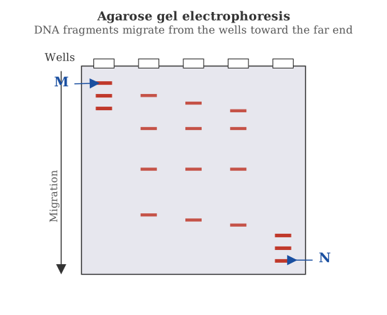
- KCET 2020Set A-11 markMCQQ.Identify the labels M and N in the following Agarose gel electrophoresis representation.
 (A) M – Digested DNA bands
(B) M – Hybridised DNA bands
(C) M – Largest DNA bands
(D) M – Smallest DNA bands
(A) M – Digested DNA bands
(B) M – Hybridised DNA bands
(C) M – Largest DNA bands
(D) M – Smallest DNA bands›Reveal solutionSolution
Agarose gel electrophoresis separates DNA fragments by size — smaller fragments travel farther. In the figure, lane M is the marker (DNA ladder) with known fragment sizes, so M shows largest DNA bands near the top and smallest near the bottom; the correct option is (C).
The concept: Why size determines distance
Agarose gel electrophoresis works like a molecular sieve. DNA is negatively charged, so when you apply an electric field across the gel, all DNA fragments move toward the positive electrode. But the agarose matrix has pores of varying sizes — smaller fragments slip through easily and travel far, while larger fragments get tangled and lag behind.
This means: distance travelled ∝ 1/(size of fragment). The largest fragments stay closest to the well (top of the gel), and the smallest fragments run farthest (bottom).
Step-by-step reasoning
-
Identify what M represents
In any gel electrophoresis figure, lane M is almost always the molecular weight marker (also called a DNA ladder). This is a mixture of DNA fragments of known, pre-determined sizes — it serves as a ruler to estimate the size of unknown fragments in other lanes.
-
Read the band pattern in lane M
The marker lane shows multiple distinct bands stacked vertically. The band nearest the well (top) is the largest fragment; the band farthest from the well (bottom) is the smallest. The bands in between correspond to intermediate sizes.
-
Match this to the options
- Option (A): "Digested DNA bands" — digestion refers to cutting DNA with restriction enzymes, which could be in sample lanes, but M is the marker, not a digested sample.
- Option (B): "Hybridised DNA bands" — hybridisation (like Southern blotting) happens after electrophoresis, not during it. The gel itself shows only size separation.
- Option (C): "Largest DNA bands" — correct in the sense that the topmost band in M is the largest, but the statement as a whole means "M shows the largest bands" (i.e., the marker contains the largest fragments). This is true relative to the sample lanes if the sample has smaller fragments. …
-
- KCET 2019Set A-11 markMCQQ.Identify the DNA sequence which can be cut using EcoRI. (A) 5'ACGAATTCAT3' 3'TGCTTAAGTA5' (B) 3'ACGAATTCAT5' 5'TGCTTAAGTA3' (C) 5'TGCTTAAGTA3' 3'ACGAATTCAT5' (D) 5'TACTTAAGCA3' 3'ATGAATTCGT5'
›Reveal solutionSolution
EcoRI recognises the palindromic sequence 5'GAATTC3' and cuts between G and A. Only option (A) contains this exact sequence on both strands in the correct antiparallel orientation.
EcoRI is a restriction endonuclease — a molecular scissors that cuts DNA at a specific recognition site. The key property of these enzymes is that they recognise palindromic sequences: the sequence on one strand, when read 5' to 3', is identical to the sequence on the complementary strand read 5' to 3'. For EcoRI, that sequence is:
5′—G A A T T C—3′
3′—C T T A A G—5′
The cut is made between the G and the first A on each strand, producing sticky ends. So to identify which option can be cut, we need to find a double-stranded DNA that contains this exact hexamer.
Let's examine each option carefully.
-
Option (A):
Top strand: 5'ACGAATTCAT3'
Bottom strand: 3'TGCTTAAGTA5'
Reading the top strand from 5' to 3', we see GAATTC starting at the third base. The bottom strand, read 5' to 3' (which means reading it from right to left as written), is 5'ATGAATTCGT3' — and that also contains GAATTC. This is the correct palindromic site. So EcoRI will cut here.
-
Option (B):
Top strand: 3'ACGAATTCAT5' — this is written in the 3' to 5' direction. To check, we must mentally flip it to 5' to 3': it becomes 5'TACTTAAGCA3'. That sequence is TACTTAAGCA — no GAATTC. The bottom strand, when flipped, is 5'ATGAATTCGT3', which does have GAATTC, but the top strand doesn't. For a restriction site to work, both strands must have the recognition sequence in the correct orientation. This fails.
-
Option (C):
Top strand: 5'TGCTTAAGTA3' — this is TGCTTAAGTA. No GAATTC. The bottom strand, read 5' to 3', is 5'TACTTAAGCA3' — also no GAATTC. So this is not a recognition site.
-
Option (D): …
-
- KCET 2018Set A-11 markMCQQ.Match the animals of Column I with the Column II and select the correct options among the following: \textbf{Column I} \hspace{2cm} \textbf{Column II} 1. DNA replication \hspace{1cm} I. RNA polymerase 2. Translation \hspace{2.5cm} II. DNA polymerase 3. Transcription \hspace{2cm} III. Reverse transcriptase 4. Reverse transcription \hspace{0.5cm} IV. Aminoacyl synthetase Select the code for the correct answer from the options given below: 1234 (A) II \hspace{0.5cm} IV \hspace{0.5cm} III \hspace{0.5cm} I (B) II \hspace{0.5cm} IV \hspace{0.5cm} I \hspace{0.5cm} III (C) II \hspace{0.5cm} III \hspace{0.5cm} IV \hspace{0.5cm} I (D) II \hspace{0.5cm} I \hspace{0.5cm} IV \hspace{0.5cm} III
›Reveal solutionSolution
Match each central-dogma process to the enzyme that catalyses it: replication→DNA polymerase, translation→aminoacyl synthetase, transcription→RNA polymerase, reverse transcription→reverse transcriptase.
Step 1 — Pair each process with its enzyme.
- DNA replication — DNA is copied into DNA. The polymerising enzyme is DNA polymerase → II.
- Translation — mRNA is decoded into protein on the ribosome. Of the enzymes listed, the one belonging to translation is aminoacyl-tRNA synthetase, which charges each tRNA with its correct amino acid (amino-acylation) → IV.
- Transcription — DNA is copied into RNA by RNA polymerase → I.
- Reverse transcription — RNA is copied into DNA (retroviruses, e.g. HIV) by reverse transcriptase (RNA-dependent DNA polymerase) → III. …
- KCET 2018Set A-11 markMCQQ.Sickle-cell anaemia is due to the following mutant gene : (A) CTC – CAC (B) CTC – GAG (C) CAC – GUG (D) GAG – GUG
›Reveal solutionSolution
A point (substitution) mutation at the 6th codon of β-globin: GAG (Glu) → GUG (Val).
Step 1 — The nature of the disease.
Sickle-cell anaemia is an autosomal recessive disorder caused by a single base substitution — a classic point mutation — in the gene for the β-globin chain of haemoglobin.
Step 2 — The molecular change.
The substitution occurs at the sixth codon of the β-globin gene, converting
GAG⟶GUG.
The middle base A is replaced by U (at the DNA level, the corresponding change is an A→T substitution).
Step 3 — The consequence. …
- KCET 2018Set A-11 markMCQQ.Which of the following sequences of mRNA are required for translation process but are not translated? (A) Stop codons (B) Anticodons (C) Sense codons (D) UTR
›Reveal solutionSolution
The 5′ and 3′ untranslated regions (UTRs) flank the coding sequence: needed for translation, never translated into protein.
Step 1 — Anatomy of a mature mRNA.
A monocistronic mRNA reads:
5′ UTR−AUG⋯coding sequence⋯stop−3′ UTR
The untranslated regions (UTRs) sit at both ends: one before the start codon and one after the stop codon.
Step 2 — Their role.
The UTRs are required for efficient translation — the 5′ UTR is where the ribosome binds and scans to find the start codon, and the 3′ UTR carries signals governing mRNA stability and localisation. But since they lie outside the reading frame (start → stop), no amino acids are made from them. They exactly satisfy 'required for translation but not translated'. ✓
Step 3 — Rule out the others. …
- KCET 2018Set A-11 markMCQQ.Identify the palindromic sequence in the following base sequences: (A) 5′−C G A T A−3′ 3′−G C T A T−5′ (B) 5′−G G A T C C−3′ 3′−C C T A G G−5′ (C) 5′−C C T G C−3′ 3′−G G A C G−5′ (D) 5′−G A A T T G−3′
›Reveal solutionSolution
Test each duplex: a palindrome reads identically 5′→3′ on both strands. Only GGATCC/CCTAGG passes.
Step 1 — The definition (and the trap).
In molecular biology a palindrome is not a word that reads the same backwards letter-by-letter. It is a duplex in which the sequence read 5′→3′ on the top strand is identical to the sequence read 5′→3′ on the bottom strand. Restriction endonucleases recognise exactly such sites — which is why the question matters.
Step 2 — Test option (B).
5′−GGATCC−3′
3′−CCTAGG−5′
Read the bottom strand in its own 5′→3′ direction, i.e. right to left: GGATCC — identical to the top strand. ✓ Palindrome. (Check complementarity too: G·C, G·C, A·T, T·A, C·G, C·G — all correct base pairs.) This is the BamHI recognition site.
Step 3 — Reject the others.
- (A) Top 5′-CGATA-3′; bottom read 5′→3′ = TATCG = CGATA. ✗ (Also 5 bp — an odd-length palindrome is impossible.)
- (C) Top 5′-CCTGC-3′; bottom read 5′→3′ = GCAGG = CCTGC. ✗ …
🎓Unlock everything free for 14 days
- ✓Full step-by-step solutions
- ✓Concept-first explanations
- ✓Methods, shortcuts & mistakes
- ✓PYQ mapping + timed mock tests
Full access for 14 days. No credit card required.