Biology · Ch 10 — Human Health and Diseases
Structure of Antibody
Structure of Antibody
Antibodies, also called immunoglobulins (Igs), are highly specific glycoproteins produced by plasma cells (themselves derived from activated B-lymphocytes) in response to antigenic stimulation — mature plasma cells can churn out antibody molecules at the extremely rapid rate of about 2,000 molecules per second.
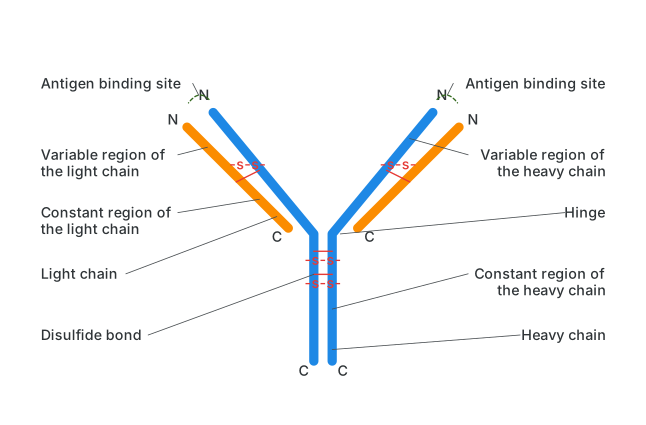
Structure : Structurally, an antibody is a 'Y'-shaped molecule built from four polypeptide chains: two identical, longer heavy (H) chains and two identical, shorter light (L) chains. These four chains are held together by disulfide bonds (-S-S-), and the region where the two arms join the stem of the 'Y' is called the hinge. Each chain — heavy or light — is itself divided into two regions: a variable region, which differs from antibody to antibody, and a constant region, which does not. The variable regions of a paired heavy and light chain together form the antigen-binding site, also called the paratope; because most antibodies have two arms, each with its own antigen-binding site, antibodies are described as bivalent. Small differences in the amino-acid sequence of the variable region are what make each antibody uniquely specific for one particular antigen.
Drawn by us to help you understand the concept clearly, and verified to make sure it's accurate. For exams, practice from your textbook's own diagram.
What this figure shows. A schematic Y-shaped antibody molecule built from four polypeptide chains — two longer heavy chains forming the stem and outer arms, and two shorter light chains paired alongside the upper arms — held together by several labelled disulfide bonds (-S-S-) at the chain junctions and at the flexible hinge region joining the stem to the two arms. Each arm's tip is labelled as an antigen-binding site, formed where the variable region of a heavy chain and the variable region of its paired light chain sit together; the remaining lower portion of each chain is labelled as its constant region. The two light chains and the upper portions of the two heavy chains are typically shown in con …
Find out
Collect information about IgG, IgA, IgM, IgD and IgE antibodies from the internet, a reference book or your teacher, and prepare a chart or a presentation. …
Drawn by us to help you understand the concept clearly, and verified to make sure it's accurate. For exams, practice from your textbook's own diagram.
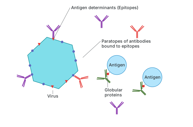
What this figure shows. A close-up schematic showing a virus particle studded with several antigen molecules, each antigen carrying a surface region labelled as an antigenic determinant (epitope); paired with it, two antibody arms are shown with their antigen-binding sites (paratopes) drawn fitting precisely against two of the virus's epitopes, illustrating the lock-and-key binding by which an antibody's paratope recognises and attaches to its matching antigenic determinant to …