Q.(a) Draw and label the T.S. of dicot leaf. OR
You're viewing a preview — the full solution, concept, methods & PYQ mapping are locked.
Start your 14-day free trial to unlock the full solution →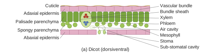
A T.S. of a typical dorsiventral (dicot) leaf shows a layered structure — upper epidermis, palisade mesophyll, spongy mesophyll (housing the vascular bundles), and lower epidermis — with stomata concentrated on the lower surface.
A hand-drawn diagram cannot be reproduced here in text, but the complete tissue arrangement to draw and label, in sequence from the adaxial (upper) to the abaxial (lower) surface, is as follows:
-
Upper epidermis (adaxial epidermis): a single layer of compactly arranged, generally colourless cells, covered externally by a thick cuticle to minimise water loss from the exposed upper surface. Stomata are few or entirely absent here in most dicot leaves.
-
Mesophyll: the photosynthetic ground tissue lying between the two epidermal layers, differentiated into two distinct zones:
a. Palisade parenchyma (palisade mesophyll): located just below the upper epidermis, made of one to three layers of elongated, columnar cells packed closely together with minimal intercellular space, oriented perpendicular to the leaf surface. These cells are densely packed with chloroplasts and are the main site of photosynthesis, since they directly face incoming light.
b. Spongy parenchyma (spongy mesophyll): located below the palisade layer, toward the lower epidermis, made of loosely arranged, irregularly shaped cells with large intercellular air spaces. It has fewer chloroplasts than the palisade layer, and its large air spaces facilitate gaseous exchange (CO2 in, O2/water vapour out) between the stomata and the photosynthesising cells.
-
Vascular bundles: embedded within the mesophyll (mainly at the spongy parenchyma level), forming the midrib centrally and lateral veins branching outward. Each vascular bundle is conjoint (xylem and phloem together), collateral (phloem only on one side of xylem) and closed (no cambium, so no further secondary growth) — with xylem oriented toward the upper (adaxial) side and phloem toward the lower (abaxial) side. The bundle is surrounded by a compact layer of parenchyma cells called the bundle sheath, which may extend to either epidermis as a bundle sheath extension.
…
Unlock everything free for 14 days
- Full step-by-step solutions
- Concept-first explanations
- Methods, shortcuts & mistakes
- PYQ mapping + timed mock tests
Full access for 14 days. No credit card required.