Question of 53
Q.Draw a diagram showing the pictorial representation of internal structure of cilia/flagella.
Rajasthan RbseRajasthan Board Senior Secondary Part-I Examination 2026Subjective· 2mImportance★★★★★
0% · 0/53 Questions
You're viewing a preview — the full solution, concept, methods & PYQ mapping are locked.
Start your 14-day free trial to unlock the full solution →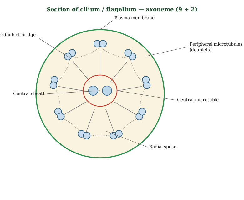
Cilia and flagella share an internal '9 + 2' arrangement of microtubules called the axoneme.
Both cilia (short, numerous) and flagella (long, usually one or two per cell) are motile, hair-like cell-surface projections with an identical internal architecture. Since a hand-drawn image cannot be included here, the structure a correct diagram should show, from the centre outward, is:
- Plasma membrane covering — the entire structure is enclosed by an extension of the cell's plasma membrane.
- Axoneme — the internal microtubule skeleton, arranged as:
- 2 central single microtubules, enclosed in a central sheath, connected by a bridge.
- 9 peripheral doublets of microtubules, arranged in a ring around the central pair, each doublet consisting of an A-tubule (complete, with dynein arms projecting toward the neighbouring doublet) and a B-tubule (incomplete, fused to the A-tubule).
- Radial spokes connect each peripheral doublet to the central sheath. …
Unlock everything free for 14 days
- Full step-by-step solutions
- Concept-first explanations
- Methods, shortcuts & mistakes
- PYQ mapping + timed mock tests
Full access for 14 days. No credit card required.