Q.(a) Explain the structure of DNA. OR
You're viewing a preview — the full solution, concept, methods & PYQ mapping are locked.
Start your 14-day free trial to unlock the full solution →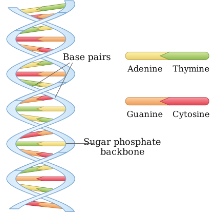
DNA, per the Watson–Crick model, is a right-handed double helix of two antiparallel nucleotide strands, joined internally by complementary, hydrogen-bonded base pairs (A–T, G–C), with a sugar-phosphate backbone on the outside.
The structure of DNA was described by James Watson and Francis Crick in 1953, based on X-ray diffraction data (chiefly from Rosalind Franklin) and Chargaff's rule of base equivalence. Its key structural features are:
-
Building block — the nucleotide: DNA is a polymer of repeating units called nucleotides. Each nucleotide has three parts: a deoxyribose sugar (a pentose sugar lacking an -OH at the 2' carbon), a phosphate group, and a nitrogenous base. The nitrogenous bases are of two types: purines (adenine, A, and guanine, G, which have a double-ring structure) and pyrimidines (cytosine, C, and thymine, T, which have a single-ring structure).
-
Polynucleotide strand: successive nucleotides are joined by phosphodiester bonds between the 3' carbon of one sugar and the 5' phosphate of the next, forming a long chain with a sugar-phosphate backbone and bases projecting sideways. Each strand therefore has a distinct polarity, with a free 5'-phosphate end and a free 3'-OH end.
-
Double helix: DNA consists of two such polynucleotide strands, wound around each other in a right-handed helix, with the sugar-phosphate backbones on the outside and the nitrogenous bases projecting inward, stacked like the rungs of a twisted ladder.
-
Antiparallel orientation: the two strands run in opposite directions — one strand runs 5'→3' while its partner runs 3'→5'.
…
Unlock everything free for 14 days
- Full step-by-step solutions
- Concept-first explanations
- Methods, shortcuts & mistakes
- PYQ mapping + timed mock tests
Full access for 14 days. No credit card required.