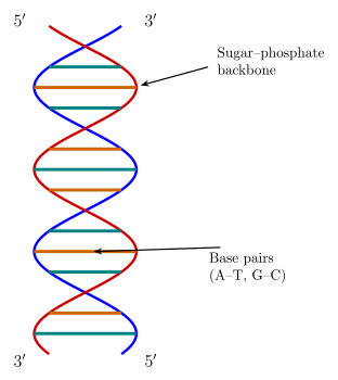
Q.a) Identify the figure given below
You're viewing a preview — the full solution, concept, methods & PYQ mapping are locked.
Start your 14-day free trial to unlock the full solution →Step 1. (a) The figure referred to is a standard DNA double helix diagram -- two antiparallel polynucleotide strands wound around each other and held together by complementary base pairing (A-T, G-C), the resting, unreplicated structure of DNA.
Step 2. (b) Redrawn as a replicating fork, the helix is shown partially unwound at one end into a Y shape: the two parental strands separate and splay apart, with each one now shown as a single strand serving as a template for a new, complementary daughter strand being synthesised alongside it.
Step 3. (c) The energy for unwinding and replication comes from the hydrolysis of high-energy phosphate bonds, chiefly ATP (and the deoxynucleotide triphosphates used as replication's own substrate, which also supply polymerisation energy); the enzyme chiefly responsible for unwinding the helix at the fork is DNA helicase, assisted by topoisomerase, which relieves the resulting torsional strain. …
Unlock everything free for 14 days
- Full step-by-step solutions
- Concept-first explanations
- Methods, shortcuts & mistakes
- PYQ mapping + timed mock tests
Full access for 14 days. No credit card required.