Q.From what you have learnt, can you tell whether enzymes are bigger or DNA is bigger in molecular size? How did you know?
Concept understanding — Enzyme Size Comparison
Enzyme Size Comparison – A First Look
Imagine you are standing in a massive warehouse. In one corner sits a single grain of rice. In another corner sits a full-sized car. Now imagine trying to find the car by looking for the grain of rice — that is the scale difference we are talking about when we compare enzymes to the molecules they work on.
Enzymes are proteins, and proteins are large, complex molecules. The substances enzymes act upon — called substrates — are usually much smaller molecules. This size difference is not just a curiosity; it is fundamental to how enzymes do their job.
The Core Idea
An enzyme is like a precision tool, and its substrate is like the raw material that fits into that tool. Because the enzyme is so much larger, it can create a specific pocket or groove — called the active site — that perfectly matches the shape of its substrate. The rest of the enzyme's bulk provides structural support, stability, and the ability to change shape slightly during the reaction.
Think of a lock and key. The lock (enzyme) is a large, complex mechanism. The key (substrate) is small and simple. The lock's internal shape is what matters — the rest of the lock is just the housing that makes the mechanism work.
Why Size Matters in Biology
The NCERT textbook emphasises that enzymes are highly specific — each enzyme typically works on only one substrate or a very small group of similar substrates. This specificity is possible because of the size difference:
- The active site is a tiny fraction of the enzyme's total structure. The rest of the enzyme acts as a scaffold, holding the active site in the correct three-dimensional shape.
- Large size allows for multiple binding sites. Some enzymes have regulatory sites where other molecules can bind and switch the enzyme on or off — like a dimmer switch on a light. A small molecule could not accommodate such complexity.
- The enzyme's bulk provides a controlled micro-environment. The active site can exclude water, create acidic or basic conditions, or apply physical stress on the substrate — all because the surrounding protein structure isolates that tiny region.
A Concrete Example from Everyday Life
Consider a scissors cutting a piece of paper. The scissors are the enzyme — large, with two blades that move precisely. The paper is the substrate — thin and flat. The scissors' size allows them to have handles, a pivot joint, and sharp edges that come together exactly. A tiny molecule-sized "scissors" could not exist because it would lack the structural parts needed to generate force and precision.
In the same way, an enzyme like amylase (found in saliva) breaks down starch into smaller sugars. The starch molecule is a long chain — but the amylase protein is many times larger, allowing it to wrap around the starch chain and snip it at specific points.
The size of an enzyme is not random. It is the result of millions of years of evolution that shaped the protein to be just large enough to create a stable, specific, and controllable active site. If enzymes were as small as their substrates, they would lack the structural complexity needed for life's chemical reactions to occur at useful speeds.
Key Points to Remember
- Enzymes are proteins — large, folded chains of amino acids.
- Substrates are small molecules that bind to the enzyme's active site.
- The enzyme's large size enables:
- A precisely shaped active site
- Multiple functional regions (active site + regulatory sites)
- A controlled chemical environment for the reaction
- This size difference is the basis of enzyme specificity — the lock-and-key or induced-fit model.
Think of it this way: a tiny screwdriver can turn a tiny screw, but the screwdriver itself needs a handle big enough for your hand to grip. The enzyme is that handle — large, stable, and designed to do one job extremely well.
Understanding why enzymes are much larger than their substrates supports the broader NCERT Class 11 Biology chapter on Biomolecules, and is often revised alongside searches like "enzyme structure and function class 11 biology" or "active site and enzyme specificity important questions." This concept underpins many NEET biochemistry questions, even though it is rarely tested as a standalone fact.
Enzymes are proteins, and DNA is a nucleic acid — both are macromolecules, but their sizes differ dramatically. A typical enzyme is made of a few hundred to a few thousand amino acids folded into a compact three-dimensional structure, giving it a molecular weight in the range of tens of thousands of daltons. DNA, on the other hand, is a polymer of millions of nucleotides arranged in a double helix that can stretch across entire chromosomes. Even a single gene (a small segment of DNA) is usually much longer than the polypeptide it codes for, because each amino acid in a protein is specified by a triplet of nucleotides (codon) in the DNA.
You can tell DNA is bigger because:
- DNA molecules in a cell are enormously long — human chromosomal DNA, if stretched out, would measure several centimeters, while enzymes are nanometer-scale globular structures.
- The molecular mass of DNA runs into millions or even billions of daltons, far exceeding that of any single enzyme.
- Functionally, DNA stores the genetic information for thousands of proteins, so it must be large enough to encode all that data.
DNA is vastly bigger than enzymes in molecular size because it is a long polymer of millions of nucleotides encoding entire genomes, whereas enzymes are compact proteins of only hundreds to thousands of amino acids.
DNA molecules are vastly larger than enzyme molecules; this follows from understanding that DNA stores the genetic instructions for making thousands of proteins (including enzymes), while each enzyme is just a single protein molecule.
When we think about molecular size in the cell, we need to remember what each molecule actually does. Enzymes are proteins—biological catalysts that speed up chemical reactions. Each enzyme is a single protein molecule, folded into a specific three-dimensional shape that allows it to bind to its substrate and carry out one particular reaction. Proteins are made of chains of amino acids, typically a few hundred to a few thousand amino acid units long. Even a large enzyme might contain around 500 to 1,000 amino acids, which translates to a molecular weight in the tens or low hundreds of thousands of daltons.
DNA, on the other hand, is the hereditary material that carries the complete genetic blueprint for an organism. A single DNA molecule in a chromosome contains millions or even billions of nucleotide base pairs strung together in a double helix. Think about what DNA has to do: it must encode the instructions for making every single protein in the organism—thousands upon thousands of different enzymes, structural proteins, regulatory proteins, and more. To store that much information, DNA molecules have to be enormously long.
The logic becomes clear when you consider the relationship between the two. DNA contains genes, and each gene is a stretch of DNA that codes for one protein. If a single gene (coding for one enzyme) might be a thousand or more nucleotides long, and a chromosome contains thousands of genes, then the entire DNA molecule must be orders of magnitude larger than any individual enzyme it encodes.
In human cells, for example, the DNA in a single chromosome can be several centimeters long when stretched out, even though it's packed tightly into the nucleus. An enzyme molecule, by contrast, is measured in nanometers—a difference of millions of times in linear dimension.
You can also reason from what you've learned about the structure of these molecules. Enzymes are compact, globular proteins. DNA is a long, thread-like polymer. The very nature of DNA as an information-storage molecule demands length; the more information to store, the longer the molecule. Enzymes, being functional units that catalyze specific reactions, don't need to be anywhere near that large.
In short, DNA molecules are far larger than enzymes. We know this because DNA must store the genetic instructions for making all the proteins in an organism (including thousands of enzymes), while each enzyme is just a single, relatively small protein molecule encoded by one gene within that vast DNA sequence.
Alternative Approach: A Quantitative "Information Content" Estimate
Instead of a purely descriptive comparison, you can reach the same conclusion by
estimating actual numbers -- a useful cross-check technique for "which is bigger"
questions in molecular biology.
Step 1: Estimate an enzyme's size.
A typical enzyme is a protein of roughly 300-500 amino acids. Since each amino acid
needs one codon (3 nucleotides) to specify it, the gene encoding a mid-sized enzyme is
only about 900-1,500 nucleotides -- and the folded protein itself occupies only a few
nanometres.
Step 2: Estimate how much DNA a genome needs to encode ALL its enzymes.
A single organism makes thousands of different enzymes (plus structural and regulatory
proteins). Even at ~1,000-1,500 nucleotides of DNA per gene, encoding thousands of genes
requires DNA millions of nucleotides long -- and that is before counting the large
non-coding regions between genes.
Step 3: Compare the two numbers directly.
One enzyme's gene: ~10^3 nucleotides. One genome: ~10^9-10^10 nucleotides (human genome
is about 3.3x10^9 base pairs, haploid). That is a difference of roughly six to seven
orders of magnitude.
Key Takeaway:
DNA must be enormously larger than any single enzyme simply because it has to store the
blueprint for every enzyme (and every other protein) an organism makes, not just one --
the size gap is a direct consequence of DNA's job as an information archive versus an
enzyme's job as a single functional catalyst.
- TG EAPCET 2025Set ap-2025-04-29-AN1 markMCQQ.Enzymes that catalyze the removal of groups from substrates by mechanism other than hydrolysis leaving double bonds are (A) Isomerases (B) Dehydrogenases (C) Hydrolases (D) Lyases
›Reveal solutionSolution
Lyases are the enzyme class that cleaves C–C, C–O, C–N, or other bonds by means other than hydrolysis or oxidation, often leaving a double bond. The correct answer is (D) Lyases.
The question tests your understanding of the six major enzyme classes defined by the International Union of Biochemistry and Molecular Biology (IUBMB). Each class is named by the type of reaction it catalyzes. The key phrase here is "removal of groups … by mechanism other than hydrolysis leaving double bonds" — that is the precise definition of a lyase.
-
Isomerases (option A) catalyze geometric or structural rearrangements within a single molecule — they don't remove groups at all. So they're out.
-
Dehydrogenases (option B) are a subclass of oxidoreductases; they remove hydrogen atoms (a type of oxidation) but do not necessarily leave a double bond as the primary product. Their mechanism involves electron transfer, not the non-hydrolytic cleavage described.
-
Hydrolases (option C) cleave bonds by adding water — that's hydrolysis. The question explicitly says "by mechanism other than hydrolysis", so hydrolases are eliminated.
-
Lyases (option D) are enzymes that catalyze the cleavage of C–C, C–O, C–N, or other bonds by elimination, leaving double bonds (or adding groups to double bonds in the reverse direction). They do not use hydrolysis or oxidation. Classic examples: fumarase (converts fumarate to malate by adding water — but in the reverse direction, it removes water leaving a double bond), aldolase (cleaves fructose-1,6-bisphosphate into two trioses), and decarboxylases (remove CO₂ leaving a double bond). This matches the description perfectly.
Watch outA common mistake is to confuse lyases with hydrolases because both break bonds. Remember: hydrolases always use water; lyases never do — they use elimination, not hydrolysis.
TipA mnemonic for the six classes: Oxidoreductases, Transferases, Hydrolases, Lyases, Isomerases, Ligases → "OTHLIL". Lyases are the fourth class, and their hallmark is forming double bonds without water.
✓Final answerThe correct option is (D) Lyases.
-
- TG EAPCET 2023Set ap-2023-05-10-FN1 markMCQQ.Which of the following enzymes belong to class 6 Ligases? (A) Glutamine synthetase (B) Hexokinase (C) Malate dehydrogenase (D) Argininosuccinase
›Reveal solutionSolution
Ligases (class 6) catalyze bond formation coupled with ATP (or similar) hydrolysis. Glutamine synthetase does exactly that — joining glutamate and ammonia using ATP — so (A) is correct.
The key to this question is understanding what defines a ligase. Enzyme classification (the EC system) groups enzymes by the type of reaction they catalyze, not by their name or the substrate alone. Class 6, ligases, are enzymes that join two molecules together by forming a new chemical bond (usually C–O, C–S, C–N, or C–C) and they always couple this bond formation to the hydrolysis of a high-energy phosphate compound — almost always ATP (or sometimes GTP, etc.). In short: ligases build bonds by burning ATP.
Now look at each option through that lens.
-
Glutamine synthetase — This enzyme catalyzes the reaction:
glutamate+NH3+ATP→glutamine+ADP+Pi
It forms a C–N bond (an amide bond) between glutamate and ammonia, and it uses ATP to drive that otherwise unfavourable reaction. That is the textbook definition of a ligase. In fact, its EC number is 6.3.1.2 — the "6" confirms it belongs to class 6.
-
Hexokinase — This transfers a phosphate group from ATP to glucose:
glucose+ATP→glucose-6-phosphate+ADP
That is a transferase (class 2), not a ligase. It moves a functional group; it does not join two separate molecules into one.
-
Malate dehydrogenase — This catalyzes:
malate+NAD+⇌oxaloacetate+NADH+H+
It is an oxidoreductase (class 1) — it transfers electrons (hydride ion) between substrates. No ATP is involved, and no new bond is formed between two molecules.
-
Argininosuccinase — This enzyme (also called argininosuccinate lyase) catalyzes:
argininosuccinate→arginine+fumarate
It breaks a bond, not forms one — it is a lyase (class 4). Lyases cleave bonds by means other than hydrolysis or oxidation, often without ATP. So it is the opposite of a ligase.
Watch outA common mistake is to think "synthetase" and "synthase" are the same. They are not. Synthetases are ligases (use ATP). Synthases are often lyases (do not use ATP). Glutamine synthetase is a ligase; but an enzyme named "glutamine synthase" would be something else. The name here is precise.
TipIf an enzyme's name ends in "-synthetase" (not just "-synthase"), it almost always signals a ligase that uses ATP. That is a quick exam shortcut — but always verify the reaction if you can.
✓Final answerThe correct option is (A) Glutamine synthetase.
-
- TG EAPCET 2023Set ap-2023-05-10-AN1 markMCQQ.Assertion (A): Enzymes are inactivated at low temperature Reason (R): Temperature changes the structure of the substrate The correct option among the following is (A) (A) and (R) are true, (R) is the correct explanation for (A) (B) (A) and (R) are true, but (R) is not the correct explanation for (A) (C) (A) is true but (R) is false (D) (A) is false but (R) is true
›Reveal solutionSolution
Enzymes become inactive at low temperature (they are temporarily switched off), so the Assertion is true. But temperature acts on the enzyme's own structure, not on the substrate, so the Reason is false — option (C).
Evaluating the Assertion
"Enzymes are inactivated at low temperature." — True.
Each enzyme works best at an optimum temperature. Well below this optimum the enzyme is temporarily inactivated: molecular motion is minimal and catalytic activity effectively stops. The enzyme is not permanently damaged and regains activity as the temperature returns towards the optimum, but at low temperature it is inactive. In the NCERT framing this statement is accepted as true.
Evaluating the Reason
"Temperature changes the structure of the substrate." — False.
Temperature acts on the enzyme, not the substrate. At high temperature the enzyme's tertiary (three-dimensional) protein structure and its active site are disrupted (denaturation); at low temperature activity simply falls. The substrate is a small molecule whose structure is not altered by these ordinary temperature changes. So the Reason is incorrect, and it cannot explain the Assertion.
Conclusion
Assertion true, Reason false.
✓Final answer(A) is true but (R) is false — option (C).
ANSWER: C
- TG EAPCET 2023Set ap-2023-05-11-AN1 markMCQQ.The essential component of many co-enzymes are (A) Vitamins (B) Metal ions (C) Hormones (D) Lipids
›Reveal solutionSolution
Co-enzymes are organic helper molecules that many enzymes require for activity, and their essential building blocks are vitamins — making option (A) the correct answer.
The question asks about the "essential component" of many co-enzymes. To answer this, you need to understand what a co-enzyme actually is and where it comes from.
An enzyme is a protein that speeds up a biochemical reaction. But many enzymes cannot work alone — they need a non-protein partner called a cofactor. Cofactors come in two types: inorganic (like metal ions) and organic (called co-enzymes). So a co-enzyme is specifically the organic type of cofactor.
Now, where do these organic co-enzymes come from? The body cannot synthesise most of them from scratch. Instead, they are derived from vitamins — the water-soluble B vitamins in particular. For example:
- Niacin (vitamin B₃) is used to make NAD⁺ and NADP⁺.
- Riboflavin (vitamin B₂) is used to make FAD and FMN.
- Thiamine (vitamin B₁) is used to make TPP (thiamine pyrophosphate).
- Pantothenic acid (vitamin B₅) is used to make coenzyme A.
So the essential component — the raw material — that the body uses to build co-enzymes is vitamins.
Let’s check the other options to be thorough:
-
Metal ions — These are indeed cofactors, but they are inorganic cofactors, not co-enzymes. Many enzymes need metal ions (e.g., Zn²⁺ in carbonic anhydrase, Mg²⁺ in kinases), but they are not the essential component of co-enzymes. Co-enzymes are organic, so metal ions are a separate category.
-
Hormones — These are chemical messengers (e.g., insulin, adrenaline) that regulate metabolism. They are not structural components of co-enzymes. Hormones and co-enzymes have completely different roles.
-
Lipids — Some lipids (like vitamin A, D, E, K) are fat-soluble vitamins, but they do not typically serve as co-enzymes. The co-enzymes are almost exclusively derived from water-soluble B vitamins. Lipids are not the essential component.
Watch outA common mistake is to confuse "cofactor" with "co-enzyme." All co-enzymes are cofactors, but not all cofactors are co-enzymes. Metal ions are cofactors, but they are not co-enzymes. The question specifically asks about the component of co-enzymes, so metal ions are not the answer.
TipA quick memory aid: The B-complex vitamins are the "building blocks" of most co-enzymes. If you memorise that NAD⁺ comes from niacin, FAD from riboflavin, and CoA from pantothenic acid, you'll never miss this type of question.
✓Final answerThe essential component of many co-enzymes is vitamins, so the correct option is (A).
- TG EAPCET 2023Set ap-2023-05-11-AN1 markMCQQ.During biochemical characterization of transforming principle by Avery, MacLeod and McCarty, the biochemicals from heat killed S cells were purified and digested with proteases, RNAase and DNAase. Digestion of DNA with DNAase enzymes shows (A) No effect of transformation (B) Inhibition of transformation (C) Delays transformation (D) Promotes transformation
›Reveal solutionSolution
Avery, MacLeod, and McCarty's experiment identified DNA as the genetic material. When DNA from heat-killed S cells was digested by DNAase, the ability to transform R cells into S cells was lost, indicating that DNA is the transforming principle. The correct option is (B).
Concept and Intuition
The question refers to a landmark experiment by Avery, MacLeod, and McCarty, which aimed to identify the chemical nature of the "transforming principle" discovered by Frederick Griffith. Griffith's experiment showed that some substance from heat-killed virulent (S) bacteria could transform non-virulent (R) bacteria into virulent (S) bacteria. However, Griffith's work did not identify what this transforming substance was.
Avery, MacLeod, and McCarty took this a step further. Their intuition was that if they could selectively destroy each major type of biochemical molecule (proteins, RNA, and DNA) from the heat-killed S cells and then test if transformation still occurred, they could pinpoint which molecule was responsible for the transformation. If destroying a particular molecule stopped transformation, then that molecule must be the transforming principle.
They used specific enzymes for this purpose:
- Proteases: Enzymes that digest and destroy proteins.
- RNAase (Ribonuclease): An enzyme that digests and destroys RNA.
- DNAase (Deoxyribonuclease): An enzyme that digests and destroys DNA.
By observing the effect of each enzyme on the transformation process, they could deduce the chemical identity of the genetic material.
Step-by-step Explanation
-
Background: Griffith's Experiment and the Transforming Principle:
Frederick Griffith, in 1928, demonstrated that a "transforming principle" existed. He observed that when live non-virulent R-strain Streptococcus pneumoniae bacteria were mixed with heat-killed virulent S-strain bacteria and injected into mice, the mice died. Live S-strain bacteria were recovered from the dead mice. This indicated that the R-strain bacteria had been transformed into the S-strain by some substance from the heat-killed S-strain.
-
Avery, MacLeod, and McCarty's Approach:
In the 1940s, Oswald Avery, Colin MacLeod, and Maclyn McCarty sought to identify the chemical nature of this transforming principle. They purified biochemicals (proteins, RNA, and DNA) from heat-killed S-strain bacteria. They then set up experiments where they would add these purified biochemicals to cultures of live R-strain bacteria.
-
Testing with Specific Enzymes:
To determine which specific molecule was the transforming principle, they treated the purified extract from heat-killed S cells with different enzymes before adding it to the R-strain bacteria:
- Treatment with Proteases: When the extract was treated with proteases (enzymes that break down proteins), transformation of R cells into S cells still occurred. This indicated that proteins were not the transforming principle.
- Treatment with RNAase: When the extract was treated with RNAase (an enzyme that breaks down RNA), transformation of R cells into S cells still occurred. This indicated that RNA was not the transforming principle.
- Treatment with DNAase: When the extract was treated with DNAase (an enzyme that specifically breaks down DNA), the transformation of R cells into S cells was inhibited. The R cells remained R cells and did not acquire the S-strain characteristics.
ImportantThe key observation was that only when DNA was destroyed by DNAase did the transforming ability cease. This directly implicated DNA as the molecule responsible for carrying the genetic information for virulence.
-
Conclusion:
The results clearly showed that the transforming principle was destroyed only when DNA was digested. This led Avery, MacLeod, and McCarty to conclude that DNA, not protein or RNA, was the genetic material responsible for heredity and transformation.
The question asks about the effect of digesting DNA with DNAase enzymes. Based on the experiment, this digestion led to the inhibition of transformation.
✓Final answerDigestion of DNA with DNAase enzymes shows (B) Inhibition of transformation.
- TG EAPCET 2023Set ap-2023-05-11-FN1 markMCQQ.Identify the incorrect combination (A) RNA polymerase I - r RNA (B) RNA polymerase II - hn RNA (C) RNA polymerase III - m RNA (D) DNA polymerase - DNA
›Reveal solutionSolution
The key is knowing which RNA polymerase transcribes which RNA in eukaryotes. RNA polymerase III transcribes tRNA, 5S rRNA, and snRNA — not mRNA. Therefore, option (C) is the incorrect combination.
The question tests your knowledge of the three eukaryotic RNA polymerases and their specific products. In prokaryotes, a single RNA polymerase handles all transcription, but eukaryotes have evolved three distinct enzymes, each dedicated to a different set of genes. This specialisation is a classic exam favourite.
The logic is straightforward: you need to match each polymerase to the RNA it synthesises. Let's check each option.
-
Option (A): RNA polymerase I → rRNA
RNA polymerase I is found in the nucleolus and transcribes the large ribosomal RNA genes (28S, 18S, and 5.8S rRNA in humans). This is correct.
-
Option (B): RNA polymerase II → hnRNA
RNA polymerase II transcribes protein-coding genes, producing heterogeneous nuclear RNA (hnRNA), which is then processed into mRNA. This is also correct.
-
Option (C): RNA polymerase III → mRNA
RNA polymerase III transcribes small, stable RNAs: tRNA, 5S rRNA, and some small nuclear RNAs (snRNA). It does not transcribe mRNA. mRNA is produced from hnRNA by RNA polymerase II. So this combination is wrong.
-
Option (D): DNA polymerase → DNA
DNA polymerase synthesises new DNA strands during replication. This is correct.
Watch outA common mistake is to confuse the product of RNA polymerase III with that of RNA polymerase II. Remember: mRNA comes from RNA pol II, not III. RNA pol III handles the "small stuff" — tRNA, 5S rRNA, snRNA.
TipA handy mnemonic: I for In the nucleolus (rRNA), II for IIn the nucleoplasm (mRNA), III for IIIny things (tRNA, 5S rRNA).
✓Final answerThe incorrect combination is (C) RNA polymerase III - mRNA.
-
- TG EAPCET 2022Set ap-2022-07-31-FN1 markMCQQ.Why RNA viruses mutate at faster rate than DNA viruses? (A) Presence of complementary strand in RNA (B) Absence of complementary strand in DNA (C) Presence of 2′–OH group in each nucleotide of DNA (D) Catalytic function of RNA
›Reveal solutionSolution
RNA viruses mutate faster than DNA viruses primarily because RNA polymerases lack the proofreading (3'→5' exonuclease) activity that DNA polymerases possess, leading to higher error rates during replication. The correct answer is (D) — the catalytic function of RNA.
Why RNA Viruses Have Higher Mutation Rates
The mutation rate during viral replication depends critically on the fidelity of the replication machinery. Let's understand why RNA viruses are the speed demons of mutation.
The Core Concept: Proofreading Makes the Difference
When genetic material is copied, errors inevitably occur. DNA polymerases have evolved sophisticated proofreading mechanisms (3'→5' exonuclease activity) that can detect and correct mismatched nucleotides immediately after they're incorporated. This is like having a spell-checker that catches typos as you type.
RNA polymerases (including RNA-dependent RNA polymerases used by RNA viruses) generally lack this proofreading ability. They're fast but sloppy copyists, incorporating incorrect nucleotides at rates 1000-10,000 times higher than DNA polymerases.
Analyzing Each Option
Let me walk through why each answer choice does or doesn't explain the mutation rate difference:
-
Option (A): "Presence of complementary strand in RNA"
- This is misleading. Both RNA and DNA can exist as single-stranded or double-stranded forms depending on the virus type. Many RNA viruses are single-stranded, but some (like reoviruses) are double-stranded. The mutation rate difference isn't fundamentally about strand complementarity.
-
Option (B): "Absence of complementary strand in DNA"
- This doesn't make sense. DNA viruses typically DO have complementary strands (double-stranded DNA). This option contains a factual error.
-
Option (C): "Presence of 2'–OH group in each nucleotide of DNA"
- This is backwards! DNA lacks the 2'–OH group (it has just H at the 2' position — hence "deoxy"). RNA has the 2'–OH group. While this chemical difference is important for stability, it's not the primary reason for mutation rate differences.
-
Option (D): "Catalytic function of RNA"
- This refers to the fact that RNA can act as both genetic material AND as a catalyst (ribozyme activity). RNA-dependent RNA polymerases are themselves proteins, but they evolved from or work with RNA-based catalytic systems. The key insight here is that RNA polymerases, which replicate RNA genomes, lack the sophisticated error-correction mechanisms of DNA polymerases. The "catalytic function" alludes to the enzymatic properties of the replication machinery.
Watch outA common misconception is that the 2'–OH group in RNA directly causes mutations. While this group does make RNA less chemically stable than DNA (more prone to hydrolysis), the mutation rate during replication is determined by the polymerase's accuracy, not the nucleotide chemistry itself.
TipThink of it this way: DNA replication is like careful manuscript copying with fact-checkers, while RNA replication is like rapid-fire transcription without editors. The trade-off for RNA viruses is that high mutation rates allow rapid adaptation to new environments and immune pressures.
The Biological Significance
The mutation rate for RNA viruses is approximately 10⁻⁴ to 10⁻⁶ substitutions per nucleotide per replication cycle, compared to 10⁻⁸ to 10⁻¹¹ for DNA-based organisms. This creates "quasi-species" populations where RNA viruses exist as swarms of related variants, enabling rapid evolution and immune evasion.
✓Final answerThe correct option is (D) — the catalytic function of RNA (and its associated replication machinery) explains why RNA viruses lack the proofreading mechanisms present in DNA replication systems, leading to higher mutation rates.
ANSWER: D
-
- TG EAPCET 2022Set ap-2022-07-31-FN1 markMCQQ.Match the following lists: List I A) \textit{E. coli} has B) Diploid human cell has C) DNA dependent RNA polymerase catalyse in D) The continuous replication is with List II I) 6.6×10⁹ bp II) 3′−5′ polarity III) 4.6×10⁶ bp IV) 5′−3′ direction The correct match is: (A) III I IV II (B) III IV II I (C) II III IV I (D) I III II IV
›Reveal solutionSolution
This question matches biological facts about genome size, DNA polarity, and replication direction. The correct pairing is: E. coli → 4.6×10⁶ bp, human diploid cell → 6.6×10⁹ bp, RNA polymerase synthesizes in 5′→3′ direction, and continuous replication occurs on the 3′→5′ template strand. The correct option is (A).
Concept & Intuition
This is a matching problem that tests your grasp of fundamental molecular biology facts:
- Genome sizes of model organisms (prokaryote vs. eukaryote).
- The directionality of nucleic acid synthesis (always 5′→3′).
- The template strand polarity for continuous (leading) replication. The trick is to recall that E. coli has a much smaller genome than a human cell, and that DNA polymerase (and RNA polymerase) always adds nucleotides to the 3′ end, so the new strand grows 5′→3′. The template strand for continuous replication is read 3′→5′.
Step-by-step reasoning
-
Match A: E. coli genome size
E. coli is a bacterium with a circular chromosome of about 4.6×10⁶ base pairs (bp). This is a standard fact.
→ A matches with III (4.6×10⁶ bp).
-
Match B: Diploid human cell genome size
A human diploid cell has about 6.6×10⁹ bp (the haploid genome is ~3.3×10⁹ bp, so diploid is double).
→ B matches with I (6.6×10⁹ bp).
-
Match C: DNA-dependent RNA polymerase catalyzes in which direction?
RNA polymerase synthesizes RNA in the 5′→3′ direction, just like DNA polymerase. It reads the template strand 3′→5′ but the new RNA chain grows 5′→3′.
→ C matches with IV (5′→3′ direction).
-
Match D: Continuous replication is with which polarity?
During DNA replication, the leading strand is synthesized continuously. The template for the leading strand is oriented 3′→5′ relative to the replication fork, so the new strand grows 5′→3′. But the question asks: "The continuous replication is with" — meaning the template strand polarity for continuous synthesis. That template is read 3′→5′.
→ D matches with II (3′→5′ polarity).
Watch outA common mistake is to think continuous replication itself has 5′→3′ polarity. But the phrase "continuous replication is with" refers to the template strand that allows continuous synthesis — that template has 3′→5′ polarity. The new strand is 5′→3′, but that's already used for RNA polymerase.
- Assemble the matches
A → III, B → I, C → IV, D → II.
This corresponds to the sequence: III, I, IV, II.
Looking at the options:
- (A) III I IV II ✓
- (B) III IV II I ✗
- (C) II III IV I ✗
- (D) I III II IV ✗
✓Final answerThe correct option is (A).
ANSWER: A
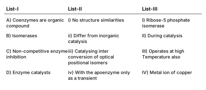
- TG EAPCET 2021Set ap-2021-08-10-FN1 markMCQQ.Match the following lists. List-I: A) Coenzymes are organic compound B) Isomerases C) Non-competitive enzyme inhibition D) Enzyme catalysts List-II:i) No structure similaritiesii) Differ from inorganic catalysisiii) Catalysing inter conversion of optical positional isomersiv) With the apoenzyme only as a transient List-III: I) Ribose-5 phosphate isomerase II) During catalysis III) Operates at high Temperature also IV) Metal ion of copper The correct match is: (A) A - iv, II; B - iii, I; C - i, IV; D - ii, III (B) A - iii, III; B - ii, I; C - iv, II; D - i, IV (C) A - iv, II; B - iii, I; C - i, III; D - ii, IV (D) A - iii, III; B - ii, IV; C - iv, I; D - i, II
›Reveal solutionSolution
Coenzymes bind the apoenzyme transiently during catalysis; isomerases (e.g. ribose-5-phosphate isomerase) interconvert isomers; non-competitive inhibitors (e.g. Cu ions) resemble the substrate not at all; and enzymes differ from inorganic catalysts, which tolerate high temperature. A–iv,II; B–iii,I; C–i,IV; D–ii,III — option (A).
The concept first
Four separate ideas are being tested at once; take them one at a time.
- Cofactors come in three flavours: prosthetic groups (tightly, permanently bound — e.g. haem in peroxidase), coenzymes (organic, loosely and transiently bound — e.g. NAD+, FAD, derived from vitamins), and metal ions (coordinated to the enzyme — e.g. Zn2+ in carboxypeptidase).
- Enzyme classes (IUB): oxidoreductases, transferases, hydrolases, lyases, isomerases, ligases.
- Inhibition: competitive — the inhibitor mimics the substrate and occupies the active site (malonate vs succinate); non-competitive — the inhibitor is structurally unlike the substrate and binds elsewhere, distorting the active site (heavy metals such as Cu2+, Hg2+, Ag+).
- Enzymes vs inorganic catalysts: enzymes are proteins — exquisitely specific, hugely efficient, but destroyed above roughly 40–50∘C. Inorganic catalysts (Pt, V2O5) are unspecific and happily operate at high temperature.
Step-by-step
- A) "Coenzymes are organic compounds." Their defining contrast with prosthetic groups is that their association with the apoenzyme is only transient → iv, and it happens during catalysis → II. So A – iv, II.
- B) Isomerases. By definition they catalyse interconversion of optical and positional isomers → iii. The stock example from the pentose-phosphate pathway is ribose-5-phosphate isomerase → I. So B – iii, I.
- C) Non-competitive inhibition. Because the inhibitor does not occupy the active site, it needs no structural similarity to the substrate → i. Textbook agents are heavy-metal ions — a metal ion of copper → IV. So C – i, IV.
- D) Enzyme catalysts. Their whole point in this list is that they differ from inorganic catalysis → ii, and the crispest way to state that difference is thermal: inorganic catalysts operate at high temperature also, while enzymes denature → III. So D – ii, III.
- Read the option. A–iv,II; B–iii,I; C–i,IV; D–ii,III is printed as option (A). Note option (C) is a decoy differing only in the last two entries (C–i,III and D–ii,IV), which would absurdly say a copper ion is the way enzymes differ from inorganic catalysts.
✓Final answerThe fully consistent matching is A–iv,II; B–iii,I; C–i,IV; D–ii,III.
ANSWER: A
🎓Unlock everything free for 14 days
- ✓Full step-by-step solutions
- ✓Concept-first explanations
- ✓Methods, shortcuts & mistakes
- ✓PYQ mapping + timed mock tests
Full access for 14 days. No credit card required.