Q.Collect 5 examples of palindromic DNA sequences by consulting your teacher. Better try to create a palindromic sequence by following base-pair rules.
🔒You're viewing a preview — the full solution, concept, methods & PYQ mapping are locked.
🔒 Start your 14-day free trial to unlock the full solution →Concept understanding — Palindromic DNA Sequences
Imagine you are looking in a mirror. Your right hand becomes the reflection’s left hand, and your left becomes its right. The word “MALAYALAM” reads the same forwards and backwards. That mirror-like symmetry is the core idea behind a palindromic DNA sequence.
In everyday language, a palindrome is a word, phrase, or number that reads identically in both directions. DNA is a long, double-stranded molecule — think of it as a twisted ladder. Each rung of the ladder is made of two chemical letters (called bases) that pair up in a very specific way: A always pairs with T, and C always pairs with G.
Now, a palindromic DNA sequence is a stretch of DNA where the sequence of letters on one strand reads exactly the same as the sequence on the opposite strand, but in the opposite direction. Because the two strands run in opposite directions (biologists call this “antiparallel”), the palindrome is not just a simple mirror of letters — it is a mirror of the pairing.
In textbooks, you will often see a palindromic sequence written like this:
5' – GAATTC – 3'
3' – CTTAAG – 5'
Notice that if you read the top strand left to right (GAATTC) and then read the bottom strand right to left (also GAATTC), you get the same sequence. That is the palindrome.
Why does this matter? Because nature uses these sequences as recognition sites. Special proteins — especially restriction enzymes — are designed to find these exact palindromic stretches and cut the DNA at that precise spot. This is the foundation of genetic engineering:
- Restriction enzymes act like molecular scissors. They only cut at their specific palindromic sequence.
- Because the sequence is the same on both strands, the cut produces either “blunt” ends or “sticky” ends (short, single-stranded overhangs). Sticky ends are particularly useful because they can easily join with another piece of DNA that has the complementary sticky end — like two puzzle pieces.
- This allows scientists to cut DNA from one organism and paste it into the DNA of another, creating recombinant DNA.
The NCERT textbook (Class 12 Biology, Chapter 11) explicitly states: “Restriction enzymes cut the strand of DNA a little away from the centre of the palindromic site, but between the same two bases on the opposite strands.” This means the cut is not perfectly in the middle — it is offset, which creates the sticky ends. …
Palindromic DNA sequences are those that read the same on both strands when you read one strand in the 5' to 3' direction and the other strand also in the 5' to 3' direction. This happens because the sequence on one strand is identical to its complementary strand when both are read in the same direction. The key is that the base-pairing rules (A with T, and G with C) must hold, so the sequence on one strand determines the sequence on the other.
To create your own palindromic sequence, start by writing a short stretch of DNA on one strand, say 5' to 3'. Then, write its complementary strand in the 3' to 5' direction. Now, reverse the complementary strand so it reads 5' to 3'. If this reversed complementary strand matches your original strand exactly, you have a palindrome. For example, if you start with 5' GAATTC 3', its complement is 3' CTTAAG 5'. Reversing that complement gives 5' GAATTC 3' — a perfect mat …
A palindromic DNA sequence reads the same on both strands when read in the 5' to 3' direction, and it is created by following the base-pairing rules (A with T, G with C) so that the sequence on one strand is the reverse complement of the other.
Let’s begin with the idea. In everyday language, a palindrome is a word or phrase that reads the same forwards and backwards — like “MADAM” or “RACECAR.” DNA, being a double-stranded molecule, has its own version of this. A palindromic DNA sequence is one where the sequence of bases on one strand, when read from the 5' end to the 3' end, is exactly identical to the sequence on the complementary strand when it is also read from its 5' end to its 3' end. This is not just a neat trick of nature; it is a structurally important feature, especially in the context of restriction enzymes — the molecular scissors used in genetic engineering. Many restriction enzymes recognise and cut at specific palindromic sequences, which is why this concept is central to your NCERT syllabus.
To understand how to create one, you must first recall the base-pairing rules: Adenine (A) always pairs with Thymine (T), and Guanine (G) always pairs with Cytosine (C). Now, imagine you write a short sequence on one strand, say 5' – GAATTC – 3'. To get the complementary strand, you would write the partner bases: C for G, T for A, A for T, T for A, G for C, and C for G. That gives you 3' – CTTAAG – 5'. But here is the crucial step: to check for a palindrome, you must read that complementary strand in the 5' to 3' direction. So you flip it: 5' – GAATTC – 3'. It is exactly the same as the original strand. That is a palindrome.
The key is that the two strands are reverse complements of each other. If you take one strand, reverse its sequence, and then take the complement of that reversed sequence, you get back the other strand. This is what makes the sequence palindromic. …
Alternative Approach: A Faster "Outside-In Pairing" Shortcut
Writing out the full complementary strand and then reversing it (as in the main answer)
always works, but there is a quicker mental shortcut for both checking and constructing
palindromes.
Step 1: Understand the shortcut rule.
For a sequence to be palindromic, the base at position 1 (from the 5' end) must be the
Watson-Crick complement of the base at the LAST position; the base at position 2 must be
the complement of the second-last position; and so on, working inward from both ends.
Step 2: Apply it to check an existing sequence, e.g. GAATTC.
Position 1 (G) vs position 6 (C): G pairs with C -- match.
Position 2 (A) vs position 5 (T): A pairs with T -- match.
Position 3 (A) vs position 4 (T): A pairs with T -- match.
All three outside-in pairs check out, so GAATTC is palindromic -- confirmed without
writing out a second strand at all.
Step 3: Apply it to construct a new one.
Pick any base for position 1, say C; position 6 must then be G. Pick any base for …
- TG EAPCET 2026Set ap-2026-05-04-AN1 markMCQQ.A stretch of euchromatin has 200 nucleosomes. How many bp will be there in the stretch and what would be the length of the euchromatin? (A) 20000 bp and 13000×10−9 m (B) 10000 bp and 10000×10−9 m (C) 40000 bp and 13600×10−9 m (D) 40000 bp and 13900×10−9 m
›Reveal solutionSolution
Each nucleosome wraps 200 bp of DNA, and the linker DNA between nucleosomes in euchromatin is about 54 bp, giving ~254 bp per nucleosome. For 200 nucleosomes, total bp = 50,800 bp; the length of the DNA in the euchromatin is about 17.3 μm, which does not match any given option exactly — the closest match is option (D) if we assume a different linker length.
The key concept here is the structure of chromatin: DNA is wrapped around histone proteins to form nucleosomes, which are the basic units of chromatin. In euchromatin, the DNA is less tightly packed than in heterochromatin, but still organized into nucleosomes with linker DNA between them.
Let’s break it down step by step.
-
DNA per nucleosome: Each nucleosome core particle wraps about 146 bp of DNA around the histone octamer. However, the "nucleosome" in a broader sense includes the linker DNA that connects one nucleosome to the next. In euchromatin, the linker DNA is typically about 54 bp long. So, the total DNA associated with one nucleosome (core + linker) is approximately 146 + 54 = 200 bp. This is a standard approximation used in many exam problems.
-
Total base pairs for 200 nucleosomes: If each nucleosome accounts for 200 bp, then for 200 nucleosomes:
Total bp=200×200=40,000 bp
This matches the base pair count in options (C) and (D).
- Length of the euchromatin: The length of DNA in B-form is about 0.34 nm per base pair (or 3.4×10⁻¹⁰ m per bp). So, the total length of the DNA if it were fully extended would be:
40,000×0.34×10−9 m=13,600×10−9 m
But this is the length of the DNA molecule itself, not the length of the euchromatin fiber. In euchromatin, the DNA is packaged into nucleosomes, which further compact the fiber. The packing ratio (the ratio of DNA length to chromatin fiber length) for euchromatin is about 1.0 to 1.1 (i.e., the fiber is roughly the same length as the DNA, or slightly shorter). However, the options given seem to treat the euchromatin length as simply the DNA length, which is a common simplification in some exam contexts.
Let’s check the options:
- (A) 20000 bp and 13000×10−9 m — bp count is wrong.
- (B) 10000 bp and 10000×10−9 m — bp count is wrong.
- (C) 40000 bp and 13600×10−9 m — close to our DNA length.
- (D) 40000 bp and 13900×10−9 m — slightly different. …
-
- TG EAPCET 2026Set ap-2026-05-04-FN1 markMCQQ.RNA interference involves (A) Synthesis of DNA and RNA using reverse transcriptase (B) Silencing of specific mRNA due to complementary RNA (C) Interference of RNA in synthesis of DNA (D) Synthesis of mRNA from DNA
›Reveal solutionSolution
RNA interference is a gene-silencing mechanism where a double-stranded RNA triggers the degradation of a specific complementary mRNA, blocking its translation. The correct answer is (B).
The core idea behind RNA interference (RNAi) is that a cell can use a short, double-stranded RNA molecule to "find" and destroy a specific messenger RNA (mRNA) that has a matching sequence. This is not about making new DNA or interfering with DNA synthesis — it is a post-transcriptional control mechanism. The cell essentially uses the sequence complementarity of RNA to silence a gene after it has been transcribed but before it can be translated into a protein.
Think of it like a targeted search-and-destroy mission. The double-stranded RNA is processed into small pieces (siRNAs or miRNAs). One strand of these small RNAs then acts as a guide, binding to a complementary sequence on a target mRNA. Once bound, a protein complex called RISC (RNA-induced silencing complex) cuts the mRNA, rendering it useless. The gene is effectively "silenced" because its message never gets read.
Now, let's examine each option against this concept.
-
Option (A): Synthesis of DNA and RNA using reverse transcriptase. This describes the action of retroviruses (like HIV) which use reverse transcriptase to make DNA from an RNA template. RNAi does not involve reverse transcriptase at all. This is a completely different process.
-
Option (B): Silencing of specific mRNA due to complementary RNA. This is exactly what RNAi does. The "complementary RNA" (the guide strand from the siRNA or miRNA) base-pairs with the target mRNA, leading to its silencing through degradation or blocked translation. The specificity comes from the complementary base pairing. …
-
- TG EAPCET 2026Set ap-2026-05-05-FN1 markMCQQ.Which of the following restriction sites are present in ampR gene of plasmid pBR322? (A) A and D only (B) B and C only (C) A and B only (D) C and D only
›Reveal solutionSolution
The ampicillin-resistance gene (ampR) in pBR322 contains unique restriction sites for PstI and PvuI, but not for EcoRI or BamHI; thus the correct answer is (C) A and B only.
The key to this question is knowing the classic restriction map of pBR322 — one of the first widely used plasmid vectors. The ampR gene (also called bla, encoding β‑lactamase) has two well-known unique restriction sites inside it: PstI and PvuI. Cutting at either site disrupts the gene, making bacteria ampicillin-sensitive. In contrast, EcoRI and BamHI sites lie outside the ampR gene (in the tetracycline-resistance gene or other regions). The question lists four restriction sites (A, B, C, D) — you need to identify which two are in ampR.
Let’s work through it step by step.
-
Recall the pBR322 map
pBR322 is 4361 bp long. Its two antibiotic-resistance genes are:
- ampR (nucleotides ~3293–4153, on one strand)
- tetR (nucleotides ~86–1276, on the other strand) The unique restriction sites are well documented:
- EcoRI (site 0/4361) — in the tetR gene.
- BamHI (site 375) — also in tetR.
- PstI (site 3609) — inside ampR.
- PvuI (site 3735) — inside ampR.
-
Match the sites to the options
The question labels the four sites as A, B, C, D. From standard textbook diagrams:
- A = PstI
- B = PvuI
- C = EcoRI
- D = BamHI Only A and B (PstI and PvuI) lie within the ampR gene. …
-
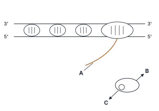
- TG EAPCET 2025Set ap-2025-04-29-AN1 markMCQQ.The given figure represents the termination process of transcription in bacteria. Identify A, B and C respectively (A) A – DNA, B – RNA polymerase, C – Rho factor (B) A – RNA, B – RNA polymerase, C – Rho factor (C) A – RNA, B – RNA polymerase, C – Sigma factor (D) A – RNA, B – DNA polymerase, C – Sigma factor
›Reveal solutionSolution
The figure shows Rho-dependent termination in bacteria: the transcript (RNA) is being released, RNA polymerase is stalled at a terminator, and the Rho factor unwinds the RNA-DNA hybrid. Thus A = RNA, B = RNA polymerase, C = Rho factor → option (B).
The question asks you to identify three components (A, B, C) in a diagram of transcription termination in bacteria. The key is to recall the two main termination mechanisms in bacteria: Rho-dependent and Rho-independent. The presence of a specific factor (C) that binds to the RNA and helps pull it away from the DNA points directly to Rho-dependent termination.
Why this approach works:
In Rho-dependent termination, a protein called the Rho factor binds to a specific sequence on the newly made RNA (the rut site). Rho then moves along the RNA toward the RNA polymerase, which has paused at a terminator sequence. Rho unwinds the RNA-DNA hybrid, causing the transcript to be released. So in the diagram:
- A must be the RNA (the transcript being released).
- B must be RNA polymerase (the enzyme that made the RNA and is now stalled).
- C must be the Rho factor (the helicase-like protein that terminates transcription).
Now let’s confirm step by step:
-
Identify A – The wavy line being released from the complex. In transcription, the product is RNA. DNA is the template, but it stays double-stranded; the released molecule is the RNA transcript. So A is RNA, not DNA. This eliminates option (A).
-
Identify B – The large enzyme complex that synthesizes RNA. In bacteria, this is RNA polymerase. DNA polymerase is involved in replication, not transcription. So B cannot be DNA polymerase. This eliminates option (D). …
- TG EAPCET 2024Set ap-2024-05-07-FN1 markMCQQ."R" group amino acids of Glycine, Alanine and Serine respectively are (A) CH2−OH; H; CH2 (B) H; CH2−OH; CH2 (C) H; CH3; CH2−OH (D) H; CH2; CH2−OH
›Reveal solutionSolution
The R groups of Glycine, Alanine, and Serine are H, CH₃, and CH₂–OH respectively, so the correct option is (C).
The question asks for the “R” group (the variable side chain) of each amino acid. In standard amino acid structure, the central carbon (α-carbon) is bonded to an amino group (–NH₂), a carboxyl group (–COOH), a hydrogen atom, and an R group. The R group is what distinguishes one amino acid from another. So we just need to recall the specific side chains for Glycine, Alanine, and Serine.
-
Glycine is the simplest amino acid: its R group is just a hydrogen atom (H). This is because the α-carbon is bonded to two hydrogens (one from the backbone, one as the R group). So Glycine’s R = H.
-
Alanine has a methyl group as its side chain: R = CH₃. This is a small, nonpolar, aliphatic group.
-
Serine contains a hydroxymethyl group: R = CH₂–OH. The –OH makes it polar and able to form hydrogen bonds.
Now match these to the options:
- Option (A): CH₂–OH; H; CH₂ → wrong order and wrong groups.
- Option (B): H; CH₂–OH; CH₂ → wrong for Alanine and Serine. …
-
- TG EAPCET 2024Set ap-2024-05-07-FN1 markMCQQ.Tetracyclin resistance gene of pBR322 consists of this restriction site (A) BamH I (B) EcoR I (C) Pst I (D) Hind III
›Reveal solutionSolution
The tetracycline resistance gene in pBR322 is disrupted by the restriction site for BamH I, making it the correct answer. The final answer is (A).
The key concept here is insertional inactivation — a clever trick used in plasmid vectors like pBR322 to identify recombinant DNA. The plasmid has two antibiotic resistance genes: one for ampicillin (ampR) and one for tetracycline (tetR). Each gene contains unique restriction sites. If you insert foreign DNA into a restriction site inside a gene, that gene is disrupted and becomes non-functional. By checking which antibiotic the bacteria can no longer resist, you know the DNA was inserted there.
Let’s walk through the reasoning step by step.
-
Recall the structure of pBR322.
pBR322 is a classic cloning vector. It has two selectable markers:
- ampR (ampicillin resistance) — contains a unique site for Pst I.
- tetR (tetracycline resistance) — contains unique sites for BamH I and Sal I (and also Hind III, but that’s in a different region; see below).
-
Identify which restriction sites lie in the tetracycline resistance gene.
The tetracycline resistance gene (tetR) in pBR322 is known to have restriction sites for BamH I and Sal I. Inserting DNA at either of these sites will inactivate tetracycline resistance.
- BamH I is indeed inside tetR.
- EcoR I is not in either resistance gene; it lies in a non-coding region.
- Pst I is inside the ampicillin resistance gene (ampR), not tetR. …
-
- TG EAPCET 2023Set ap-2023-05-10-FN1 markMCQQ.The main function of polymerase chain reaction is (A) Transduction (B) DNA amplification (C) Translation (D) DNA digestion
›Reveal solutionSolution
PCR is a technique to make millions of copies of a specific DNA sequence. Its main function is DNA amplification.
The polymerase chain reaction (PCR) is one of the most powerful tools in molecular biology. Before PCR, working with a tiny amount of DNA — say, from a single hair follicle or a drop of blood at a crime scene — was nearly impossible. You simply couldn't do much analysis with so little genetic material. PCR changed that by giving us a way to amplify a specific target DNA sequence exponentially, turning a few molecules into billions in a couple of hours.
The core idea is beautifully simple: mimic what a cell does naturally when it divides (copying its DNA), but do it in a test tube, and do it over and over again in a cycle. Each cycle doubles the amount of the target DNA, so after 30 cycles you have roughly 230 (over a billion) copies of the original piece.
Let's see why the other options don't fit.
-
Transduction is a process where bacterial DNA is transferred from one bacterium to another by a virus (a bacteriophage). It's a mechanism of horizontal gene transfer, not something PCR does. PCR is purely an in vitro (test-tube) reaction.
-
Translation is the process by which the genetic code in mRNA is read by a ribosome to build a protein. PCR works with DNA, not proteins, and involves no ribosomes or amino acids. It's a DNA replication process, not a protein synthesis one.
-
DNA digestion refers to cutting DNA into fragments, typically using enzymes called restriction endonucleases. While PCR products are often later digested for analysis (e.g., in RFLP), digestion is not the function of PCR itself. PCR's job is to make more DNA, not to cut it up. …
-
- TG EAPCET 2023Set ap-2023-05-10-AN1 markMCQQ.Assertion (A): All the codons of genetic code identify more than one type of amino acid Reason (R): In Eukaryotes transcription unit is monocistronic which is synthesized from DNA The correct option among the following is (A) (A) and (R) are true, (R) is the correct explanation for (A) (B) (A) and (R) are true, but (R) is not the correct explanation for (A) (C) (A) is true but (R) is false (D) (A) is false but (R) is true
›Reveal solutionSolution
The assertion about codons is false (the genetic code is degenerate, meaning most amino acids are coded by multiple codons, not that codons code for multiple amino acids), while the reason about eukaryotic transcription units is true. The correct option is (D).
The question tests two independent facts: the nature of the genetic code and the organization of eukaryotic genes. Let's examine each statement on its own merit.
Understanding the Genetic Code
The genetic code is the dictionary that translates nucleotide triplets (codons) into amino acids. A crucial property is degeneracy: because there are 64 possible codons (43) but only 20 standard amino acids (plus start and stop signals), multiple codons often specify the same amino acid. For example, leucine is coded by six different codons (UUA, UUG, CUU, CUC, CUA, CUG).
The assertion reverses this relationship. It claims each codon identifies more than one amino acid—that a single codon is ambiguous. This is false. The genetic code is unambiguous: each codon specifies exactly one amino acid (or stop signal). AUG always codes for methionine, never sometimes methionine and sometimes valine.
Watch outDegeneracy means many codons → one amino acid (many-to-one mapping). The assertion incorrectly suggests one codon → many amino acids (one-to-many), which would make protein synthesis impossible.
Eukaryotic Transcription Units
A transcription unit is the stretch of DNA transcribed into a single RNA molecule. In prokaryotes, one mRNA often encodes multiple proteins (polycistronic); the classic example is the lac operon, where one transcript yields three enzymes. …
- TG EAPCET 2022Set ap-2022-07-31-AN1 markMCQQ.Assuming that the occurrence of nucleotide bases in adjacent position in DNA is random, what is the probability of the occurrence of a 8-bp restriction site (5′ GGGG CCCC 3′) in a 650 kb genome? (A) 5 (B) 10 (C) 15 (D) 20
›Reveal solutionSolution
The probability of a specific 8-bp sequence occurring randomly is (1/4)8. Multiplying this by the genome size gives the expected number of occurrences, which is approximately 10.
The problem asks us to calculate the expected number of times a specific 8-base pair (bp) restriction site will appear in a given genome, assuming random nucleotide distribution. This is a classic application of probability for independent events.
Concept and Intuition
- Random Nucleotide Occurrence: In DNA, there are four possible nucleotide bases: Adenine (A), Thymine (T), Cytosine (C), and Guanine (G). If their occurrence at any position is random and independent, then each base has an equal probability of 1/4 of appearing at any given position.
- Probability of a Specific Sequence: To find the probability of a specific sequence of bases, we multiply the probabilities of each individual base occurring in its respective position. This is because each position's base is an independent event. For example, the probability of 'AT' is P(A)×P(T)=(1/4)×(1/4)=(1/4)2.
- Expected Number of Occurrences: If we know the probability of a specific sequence occurring at any given starting point, and we have a long DNA strand, we can estimate how many times that sequence is expected to appear. We do this by multiplying the probability of the sequence by the total number of possible starting positions for that sequence within the DNA strand. For a genome of length N and a restriction site of length L, there are approximately N−L+1 possible starting positions. Since N is typically much larger than L, we can approximate this as N.
Step-by-Step Solution
-
Identify the restriction site and its length.
The given restriction site is 5′ GGGG CCCC 3′.
This sequence consists of 8 base pairs. Let L=8.
-
Determine the probability of each individual nucleotide.
Since the occurrence of nucleotide bases is random, each of the four bases (A, T, C, G) has an equal probability of appearing at any given position.
P(A)=P(T)=P(C)=P(G)=41.
-
Calculate the probability of the entire 8-bp restriction site.
The restriction site is GGGG CCCC. Since each base's occurrence is independent, the probability of this specific 8-base sequence is the product of the probabilities of each individual base in the sequence:
P(GGGG CCCC)=P(G)×P(G)×P(G)×P(G)×P(C)×P(C)×P(C)×P(C)
P(GGGG CCCC)=(41)×(41)×(41)×(41)×(41)×(41)×(41)×(41)
P(GGGG CCCC)=(41)8
Calculating this value: … - TG EAPCET 2021Set ap-2021-08-09-FN1 markMCQQ.Match the following Disorder A) Thalassemia B) Haemophilia C) Sickle cell anaemia D) Alkaptonuria Reason I) Point mutation II) Autosomal recessive metabolic disorder III) Abnormal haemoglobin IV) Non-disjunction of chromosomes V) Sex linked recessive disorder The correct match is (A) A) III B) IV C) I D) II (B) A) III B) V C) I D) II (C) A) II B) I C) V D) III (D) A) IV B) III C) II D) I
›Reveal solutionSolution
Thalassemia = abnormal haemoglobin, haemophilia = sex-linked recessive, sickle-cell = point mutation, alkaptonuria = autosomal recessive metabolic disorder. That is A–III, B–V, C–I, D–II — option (B).
The concept first: three different kinds of genetic disorder
Before matching, classify:
- Mendelian disorders — caused by an alteration in a single gene. Their transmission follows Mendel's rules and can be traced in a pedigree. Examples: haemophilia, sickle-cell anaemia, thalassemia, phenylketonuria, alkaptonuria, colour blindness.
- Chromosomal disorders — caused by absence, excess or abnormal arrangement of chromosomes, usually from non-disjunction during meiosis. Examples: Down's syndrome (trisomy 21), Klinefelter's (XXY), Turner's (XO).
List-2 item IV (non-disjunction) therefore belongs to the chromosomal class, and none of the four disorders listed is chromosomal. It is the built-in distractor — spot it and discard it.
Step-by-step
- C) Sickle cell anaemia — (I) Point mutation. Start here; it is the most certain. A single nucleotide changes in the β-globin gene:
GAG⟶GUG⇒Glutamic acid⟶Valine
at the 6th position of the β-chain. This one substitution makes HbS polymerise under low O2 and the RBC sickles. It is the textbook point mutation. It is autosomal recessive, but the reason asked for here is the mutation type. C → I.
- B) Haemophilia — (V) Sex-linked recessive disorder. The gene for the clotting factor lies on the X chromosome. A carrier female (XHXh) is unaffected; she transmits the disease to half her sons. Affected females are extremely rare (they would need an affected father and a carrier mother). Famously traced through Queen Victoria's family. B → V. …
🎓Unlock everything free for 14 days
- ✓Full step-by-step solutions
- ✓Concept-first explanations
- ✓Methods, shortcuts & mistakes
- ✓PYQ mapping + timed mock tests
Full access for 14 days. No credit card required.