Q.In some viruses, DNA is synthesised by using RNA as template. Such a DNA is called:
🔒You're viewing a preview — the full solution, concept, methods & PYQ mapping are locked.
🔒 Start your 14-day free trial to unlock the full solution →Concept understanding — Types Of Polymerases
You have probably never heard the word "polymerase" before, but you already understand what it does. Think of a polymer as a long chain — like a necklace made of many identical or similar beads. In biology, the most famous polymers are DNA and RNA, which are chains of smaller units called nucleotides. A polymerase is simply the enzyme (a protein machine) that links those beads together to build the chain.
In the context of your syllabus, the term "polymerases" almost always refers to DNA polymerases and RNA polymerases. These are the two main types you need to know. They are not interchangeable; each has a specific job and a specific set of rules.
1. DNA Polymerase — The Copyist
DNA polymerase is the enzyme responsible for DNA replication — making an exact copy of the entire DNA molecule before a cell divides. Imagine you have a master blueprint (the original DNA). DNA polymerase reads that blueprint and builds a matching second copy.
Key features to remember:
- It can only add new nucleotides to an existing strand (it needs a "primer" to start).
- It works in one direction only: from the 5' end to the 3' end of the new strand.
- It has a proofreading ability. If it accidentally puts the wrong bead on the chain, it can cut it out and replace it. This is why DNA replication is so accurate.
In NCERT, you will encounter three types of DNA polymerases in prokaryotes (like E. coli): DNA polymerase I, II, and III. Of these, DNA polymerase III is the main enzyme that does the bulk of replication. DNA polymerase I helps remove the RNA primer and fill in gaps. DNA polymerase II is mainly involved in repair.
2. RNA Polymerase — The Transcriber
RNA polymerase does a different job. It is responsible for transcription — copying a specific gene from DNA into a messenger RNA (mRNA) molecule. Think of it as a photocopier that makes a working copy of just one page of the blueprint, not the whole book.
Key differences from DNA polymerase:
- It does not need a primer to start. It can begin building the RNA chain from scratch.
- It uses DNA as a template but builds an RNA strand (which uses uracil instead of thymine).
- It is less accurate than DNA polymerase — it has no proofreading ability. This is acceptable because RNA molecules are temporary and get replaced. …
The key here is the template. When a virus uses RNA as a template to make DNA, the process is reverse transcription, and the DNA produced is complementary to the original RNA.
- cDNA stands for complementary DNA. It is synthesised from an RNA template using the enzyme reverse transcriptase. This is exactly what happens in retroviruses like HIV.
- A-DNA and B-DNA are just different structural forms (conformations) of the DNA double helix — they have nothing to do with the template used for synthesis. …
When a virus uses RNA as a template to make DNA, that DNA is called complementary DNA (cDNA).
The question touches on a fascinating exception to the central dogma of molecular biology — the flow of genetic information. Normally, DNA makes RNA, and RNA makes protein. But some viruses, most famously retroviruses like HIV, reverse this flow. They carry their genetic information as RNA, and once inside a host cell, they use that RNA as a template to synthesise DNA.
This process is catalysed by an enzyme called reverse transcriptase. The DNA produced in this way is not identical to any pre-existing DNA in the cell. Instead, it is a complementary copy of the viral RNA. That is why it is called complementary DNA, or cDNA.
The term "complementary" here means that the DNA sequence is exactly complementary to the RNA template — each RNA base pairs with its DNA counterpart (A with T, U with A, G with C, and C with G).
Now, let us look at the options given:
-
A-DNA and B-DNA are structural forms of DNA — different helical conformations that DNA can adopt under certain conditions. They have nothing to do with the template used for synthesis. A-DNA is a right-handed helix that is shorter and wider than the more common B-DNA, but both are simply physical shapes of the DNA molecule.
-
rDNA stands for recombinant DNA — DNA that has been artificially created by combining genetic material from different sources. This is a tool used in biotechnology, not a natural product of viral replication. …
Decode what each answer's own name implies before considering the biology: 'A-DNA' and 'B-DNA' name helical conformations — shape variants of ordinary double-stranded DNA — not a synthesis route. 'rDNA' names DNA assembled in vitro from multiple sources (recombinant DNA), a laboratory construction technique, not a natural viral product. Only 'cDNA' names …
- TG EAPCET 2026Set ap-2026-05-04-AN1 markMCQQ.Which of the following is wrongly matched pair (A) Repressor → Protein coded by regulatory gene of operon (B) Operon → Promotor, Structural genes, operator (C) Translation → Polymerisation of amino acids and polypeptide chain formation (D) Transcription → Attachment of amino acid to a specific t-RNA
›Reveal solutionSolution
The question asks to identify the incorrectly matched pair among common molecular biology terms. The process of attaching an amino acid to its specific tRNA is called aminoacylation, not transcription. Thus, option (D) is the wrongly matched pair.
Understanding the fundamental processes of gene expression – transcription and translation – along with the concept of an operon, is crucial for answering this question. Each option describes a key component or process in molecular biology. We need to evaluate if the description accurately matches the term.
-
Analyze Option (A): Repressor → Protein coded by regulatory gene of operon
- In an operon system (like the lac operon in bacteria), a regulatory gene (often denoted as the i gene) is responsible for synthesizing a repressor protein.
- This repressor protein then binds to the operator region of the operon, thereby regulating the transcription of the structural genes.
- Therefore, this statement is correct.
-
Analyze Option (B): Operon → Promotor, Structural genes, operator
- An operon is a functional unit of DNA containing a cluster of genes under the control of a single promoter.
- It typically consists of:
- A promoter region: where RNA polymerase binds to initiate transcription.
- An operator region: a segment of DNA to which a repressor protein binds, regulating gene expression.
- Structural genes: genes that code for proteins involved in a particular metabolic pathway.
- These three components are indeed the core parts of an operon.
- Therefore, this statement is correct.
-
Analyze Option (C): Translation → Polymerisation of amino acids and polypeptide chain formation
- Translation is the biological process where messenger RNA (mRNA) is decoded by a ribosome to produce a specific amino acid chain, or polypeptide.
- This process involves the sequential addition of amino acids, linked by peptide bonds, to form a growing polypeptide chain. This is precisely the "polymerisation of amino acids and polypeptide chain formation."
- Therefore, this statement is correct.
-
Analyze Option (D): Transcription → Attachment of amino acid to a specific t-RNA …
-
- TG EAPCET 2026Set ap-2026-05-04-AN1 markMCQQ.2 A + 2 G–3P Transketolase 2 xylulose–5–Phosphate + 2 B In the above reaction A and B are respectively (A) Sedoheptulose-7-phosphate and erythrose-4-phosphate (B) Fructose-6-phosphate and ribose 5-phosphate (C) Fructose-6-phosphate and erythrose-4-phosphate (D) DHAP and fructose-6-phosphate
›Reveal solutionSolution
Transketolase transfers a two-carbon unit from a ketose donor to an aldose acceptor. Dividing the given equation by 2, it reads: (ketose A) + glyceraldehyde-3-phosphate → xylulose-5-phosphate + (aldose B). Carbon counting identifies A = fructose-6-phosphate and B = erythrose-4-phosphate — option (C).
Transketolase always moves a two-carbon "ketol" fragment (−CO−CH2OH) from a ketose sugar to an aldose sugar, converting the ketose donor into an aldose 2 carbons shorter, and the aldose acceptor into a ketose 2 carbons longer.
The given equation, 2A+2 G-3PTransketolase2 xylulose-5-P+2B, halves to:
A+G-3P→xylulose-5-P+B.
Glyceraldehyde-3-phosphate (G–3P) is a 3-carbon aldose. As the acceptor, it gains 2 carbons to become the 5-carbon ketose xylulose-5-phosphate (3+2=5) — consistent with the given product.
So A must be the donor ketose, losing 2 carbons to become the aldose B. In the pentose phosphate pathway, the transketolase reaction that uses glyceraldehyde-3-phosphate as the acceptor and produces xylulose-5-phosphate is:
Fructose-6-phosphate (6C ketose)+G-3P→Xylulose-5-P (5C ketose)+Erythrose-4-phosphate (4C aldose). …
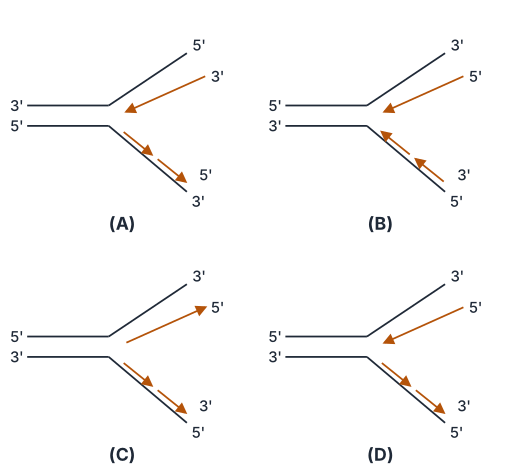
- TG EAPCET 2025Set ap-2025-04-30-FN1 markMCQQ.Which of the following figures correctly shows the replication fork formed during DNA replication? (A) [FIGURE] Replication fork diagram — upper template labelled 3' at its left end, lower template labelled 5' at its left end, with the new strands' 5'/3' ends and synthesis arrows marked at the fork (B) [FIGURE] Replication fork diagram — upper template labelled 5' at its left end (new strand ends 3' above 5'), lower template labelled 3' at its left end, arrows pointing away from the fork on the lower strand (C) [FIGURE] Replication fork diagram — upper template labelled 5' at its left end with the new-strand arrow pointing towards the fork (up-right), lower template labelled 3' at its left end (D) [FIGURE] Replication fork diagram — upper template labelled 5' at its left end with the new strand running 3'/5' and its arrow pointing back toward the fork, lower template labelled 3' at its left end with discontinuous-synthesis arrows
›Reveal solutionSolution
A valid replication-fork diagram must obey two constraints — antiparallel templates and 5′→3′ synthesis only — which force one arrow to point towards the fork (leading) and the other away from it (lagging). The figure in option (D) is the only one that is self-consistent.
The concept first: why the fork looks lop-sided
Rule 1 — DNA polymerase has only one direction. The enzyme adds a nucleotide by attacking the incoming dNTP with the free 3′-OH of the growing chain. It has no chemistry for adding to a 5′ end. Therefore:
every new strand grows 5′⟶3′
and, since the new strand is antiparallel to its template, the enzyme reads its template 3′→5′.
Rule 2 — the two parental strands are antiparallel. In the duplex, one strand runs 5′→3′ left-to-right and the other 3′→5′ left-to-right. So when helicase opens the fork, the two exposed templates point in opposite directions relative to the moving fork.
The consequence. Consider the direction the fork travels (into the unopened duplex):
- On the template whose 3′ end lies toward the fork, reading 3′→5′ means moving in the same direction as the fork. The polymerase can therefore chase the fork continuously ⇒ this is the leading strand, synthesised in one unbroken piece, its arrow pointing towards the fork.
- On the other template the polarity is reversed. Reading 3′→5′ now means moving away from the fork. The polymerase must wait for a stretch of template to be exposed, then back-track and synthesise a short piece away from the fork; repeat. ⇒ this is the lagging strand, made as Okazaki fragments later joined by DNA ligase, with arrows pointing away from the fork.
So the signature of a correct fork diagram is asymmetry: one arrow in, one arrow out.
Step-by-step: audit the four figures
A useful two-part checklist for each option — (i) do the arrows show one strand made towards and one away from the fork? (ii) are those directions consistent with the printed 3′/5′ polarity labels? …
- TG EAPCET 2024Set ap-2024-05-07-AN1 markMCQQ.Match the following List - I A Operator site B Promotor site C Regulator gene D Structural gene List - II I Binding site for RNA polymerase II Binding site for repressor III Codes for Enzyme IV Codes for repressor (A) A-I, B-IV, C-II, D-III (B) A-I, B-II, C-IV, D-III (C) A-II, B-I, C-III, D-IV (D) A-II, B-I, C-IV, D-III
›Reveal solutionSolution
The lac operon regulates gene expression in bacteria. The operator site binds the repressor, the promoter site binds RNA polymerase, the regulator gene codes for the repressor, and structural genes code for enzymes. The correct match is (D).
The lac operon is a classic example of gene regulation in prokaryotes, specifically in E. coli. It controls the expression of genes necessary for the metabolism of lactose. Understanding the function of each component — the promoter, operator, regulator gene, and structural genes — is crucial to grasp how gene expression is turned on or off in response to environmental cues.
Here's how the components match their functions:
-
A. Operator site:
The operator site is a specific DNA sequence located immediately downstream of the promoter and upstream of the structural genes. Its primary role is to act as a switch for transcription. It serves as the binding site for the repressor protein. When the repressor protein binds to the operator, it physically obstructs the movement of RNA polymerase, preventing it from transcribing the structural genes. This effectively turns off gene expression.
Therefore, A matches with II (Binding site for repressor).
-
B. Promoter site:
The promoter site is a DNA sequence located upstream of the operator and structural genes. It is the recognition and binding site for RNA polymerase. RNA polymerase must bind to the promoter to initiate the transcription of the genes that follow. Without a functional promoter, transcription cannot begin.
Therefore, B matches with I (Binding site for RNA polymerase).
-
C. Regulator gene (or i gene): …
-
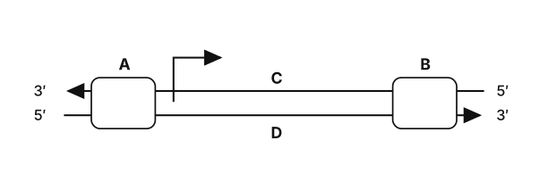
- TG EAPCET 2024Set ap-2024-05-07-FN1 markMCQQ.Given is the schematic structure of the transcription unit. Select the correct answer regarding A, B, C and D. [FIGURE: a double-stranded DNA transcription unit; the upper strand runs 3' (left) to 5' (right) and the lower strand 5' (left) to 3' (right). Box A lies at the left end of the unit, box B at the right end, C labels the upper strand and D labels the lower strand; an arrow at the right of box A shows the direction of transcription.] (A) A - Terminator, B - Promotor, C - Template strand, D - Coding strand (B) A - Promotor, B - Terminator, C - Coding strand, D - Template strand (C) A - Promotor, B - Terminator, C - Template strand, D - Coding strand (D) A - Terminator, B - Promotor, C - Coding strand, D - Template strand
›Reveal solutionSolution
The bent arrow next to box A marks the transcription start, so A = promoter and B = terminator. The upper strand runs 3′→5′ along the direction of transcription, making it the template (C), and the lower 5′→3′ strand is the coding strand (D). Option (C).
The concept first — what a transcription unit is made of
A transcription unit has three components:
- Promoter — the DNA sequence, located upstream (towards the 5' end of the coding strand), where RNA polymerase binds. It defines the template strand.
- The structural gene — the stretch that is actually copied.
- Terminator — located downstream (towards the 3' end of the coding strand), where transcription stops.
And the two strands have fixed names:
- Template (antisense/minus) strand — the one the polymerase reads. Since RNA is built 5′→3′ and DNA–RNA pairing is antiparallel, this strand must have 3′→5′ polarity in the direction of transcription.
- Coding (sense/plus) strand — the other one. It has 5′→3′ polarity and its base sequence is identical to the mRNA (except T instead of U). It is not copied at all; it is called "coding" only because it reads like the message.
Step-by-step, using the figure
- Find the direction of transcription. The bent arrow, which marks the start point and points rightwards, sits immediately to the right of box A. RNA polymerase always begins at the promoter, so:
A=Promoter
- The far end must therefore be the stop signal: B=Terminator …
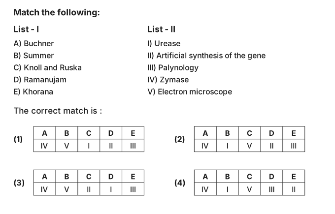
- TG EAPCET 2022Set ap-2022-07-31-AN1 markMCQQ.Match the following: List-I: A) Buchner B) Summer C) Knoll and Ruska D) Ramanujam E) Khorana List-II: I) Urease II) Artificial synthesis of the gene III) Palynology IV) Zymase V) Electron microscope The correct match is: (A) A - IV; B - V; C - I; D - II; E - III (B) A - IV; B - I; C - V; D - II; E - III (C) A - IV; B - V; C - II; D - I; E - III (D) A - IV; B - I; C - V; D - III; E - II
›Reveal solutionSolution
Buchner → zymase; Sumner → urease; Knoll & Ruska → electron microscope; Ramanujam → palynology; Khorana → artificial gene synthesis. That is A–IV, B–I, C–V, D–III, E–II — option (D).
The concept first
This is a straight history-of-biology recall item. The safe strategy is to anchor the two or three you are certain about, then let the options eliminate one another.
Step-by-step
- A) Buchner → IV (Zymase). In 1897 Eduard Buchner ground yeast cells and showed that the resulting cell-free extract could still ferment sugar to alcohol. He named the active principle zymase. This was a landmark: it proved fermentation is a chemical process catalysed by a substance, not a mystical property of living cells. Nobel Prize, 1907.
- B) Sumner → I (Urease). In 1926 James B. Sumner crystallised urease from jack bean — the first enzyme ever obtained in pure crystalline form — and demonstrated that it was a protein. This settled a long-running dispute about the chemical nature of enzymes. Nobel Prize, 1946.
- C) Knoll and Ruska → V (Electron microscope). Max Knoll and Ernst Ruska built the first transmission electron microscope in 1931–32, breaking the resolution limit imposed on light microscopy by the wavelength of visible light. Nearly everything we know about cell ultrastructure — ER, ribosomes, membranes — flowed from this instrument. Ruska received the Nobel Prize in 1986. …
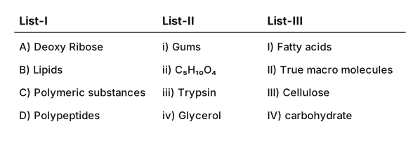
- TG EAPCET 2021Set ap-2021-08-09-AN1 markMCQQ.Match the following lists. List-I A) Deoxy Ribose B) Lipids C) Polymeric substances D) Polypeptides List-IIi) Gumsii) C5H10O4iii) Trypsiniv) Glycerol List-III I) Fatty acids II) True macro molecules III) Cellulose IV) carbohydrate The correct match is: (A) A) ii, IV B) iii, I C) i, III D) iv, II (B) A) iii, II B) ii, I C) i, III D) iv, IV (C) A) iv, I B) i, III C) iii, IV D) ii, II (D) A) ii, IV B) iv, I C) i, III D) iii, II
›Reveal solutionSolution
Deoxyribose is the C5H10O4 carbohydrate; lipids give glycerol and fatty acids; gums/cellulose are the polymeric substances; and polypeptides such as trypsin are the true macromolecules. That is A–ii,IV · B–iv,I · C–i,III · D–iii,II — option (D).
The concept first
Biomolecules split cleanly along three axes, and this question tests all three at once: the formula (List-II sometimes), the hydrolysis product/example (List-II), and the chemical class or status (List-III). Anchor on the item you know with certainty and let the rest follow.
Step-by-step
-
A) Deoxyribose. Ribose is C5H10O5. "Deoxy" means one oxygen fewer at C-2, so deoxyribose is C5H10O4 ⇒ List-II (ii). It is a pentose sugar, i.e. a carbohydrate ⇒ List-III (IV). A → ii, IV.
-
B) Lipids. A triglyceride hydrolyses as
Fat+3H2O⟶Glycerol+3Fatty acids
So lipids are built from glycerol ⇒ (iv) and fatty acids ⇒ (I). B → iv, I.
- C) Polymeric substances. Gums (i) are polysaccharide polymers, and cellulose (III) is the archetypal plant polymer — a β-1,4 linked glucose homopolymer. C → i, III. …
-
🎓Unlock everything free for 14 days
- ✓Full step-by-step solutions
- ✓Concept-first explanations
- ✓Methods, shortcuts & mistakes
- ✓PYQ mapping + timed mock tests
Full access for 14 days. No credit card required.