Q.Discuss the process of translation in detail.
🔒You're viewing a preview — the full solution, concept, methods & PYQ mapping are locked.
🔒 Start your 14-day free trial to unlock the full solution →Concept understanding — Gene Expression Regulation
Imagine a library with thousands of books. Every cell in your body has the same library — the same complete set of genes in your DNA. But a skin cell does not need to read the book on "how to make stomach acid," and a stomach cell does not need the book on "how to make skin pigment." If every cell tried to read every book at once, the library would be chaos. Gene expression regulation is the system that decides which books are opened, which are kept closed, and when to put a book back on the shelf.
At its simplest, gene expression is the process by which information from a gene is used to make a functional product — usually a protein. Regulation means this process is not automatic; it is controlled. Cells turn genes on or off, or adjust how much product is made, depending on what the body needs at that moment.
Why does this matter? Without regulation, every cell would be identical and useless. Regulation is what makes a muscle cell different from a nerve cell, even though both contain the same DNA. It also allows your body to respond to changes — like producing more red blood cells when you move to high altitude, or repairing damage after a cut.
Think of gene regulation like a dimmer switch, not just an on/off button. Some genes are turned up high, some are turned down low, and many are in between. This fine-tuning is essential for health.
The NCERT textbook explains that regulation can happen at several stages. The most important stage in bacteria (like E. coli) is at the start of transcription — when the gene is first copied into RNA. In higher organisms, regulation is more complex and can occur at multiple points: before transcription, during RNA processing, during translation (making protein), and even after the protein is made.
Key points to remember:
- All cells have the same DNA, but different sets of genes are active in different cells.
- Regulation is dynamic — genes can be turned on and off in response to signals from inside or outside the cell.
- Mistakes in regulation can lead to diseases like cancer, where genes that should be off stay on, or genes that should be on stay off. …
Translation is the process by which the sequence of codons on mRNA is decoded into a sequence of amino acids to form a polypeptide chain. It occurs on ribosomes in the cytoplasm. The key players are mRNA (the template), tRNA (which brings specific amino acids and has an anticodon that pairs with the mRNA codon), aminoacyl-tRNA synthetases (which charge tRNAs with their correct amino acids), and the ribosome (composed of large and small subunits).
The process has three main stages:
-
Initiation: The small ribosomal subunit binds to the mRNA near the start codon (AUG). The initiator tRNA, carrying methionine, pairs with the start codon. Then the large ribosomal subunit joins, forming a functional ribosome with two sites: the A site (aminoacyl) and the P site (peptidyl).
-
Elongation: A charged tRNA enters the A site, its anticodon pairing with the next codon. A peptide bond forms between the amino acid in the P site and the new amino acid in the A site, catalyzed by peptidyl transferase (an activity of the ribosomal RNA). The ribosome then translocates one codon forward, moving the now-uncharged tRNA to the E site (exit) and the peptidyl-tRNA to the P site, freeing the A site for the next tRNA. …
Translation is the process by which the genetic information carried by mRNA is decoded to produce a specific sequence of amino acids, forming a polypeptide chain.
To understand translation, you first need to see it as the final act in the drama of gene expression. After transcription has produced a messenger RNA (mRNA) copy of a gene, that mRNA must now be "read" to build a protein. This reading happens on a molecular machine called the ribosome, and the language being translated is the triplet code of codons on the mRNA into the language of amino acids.
The process requires three main players: mRNA (the template), tRNA (the adapter that brings the correct amino acid), and the ribosome (the factory). Each tRNA molecule has a specific anticodon that base-pairs with a complementary codon on the mRNA, and it carries the corresponding amino acid at its other end. The ribosome itself is composed of two subunits — a large subunit and a small subunit — that come together only when translation begins.
Initiation is the first phase. In prokaryotes, the small ribosomal subunit binds to the mRNA at a specific sequence called the Shine-Dalgarno sequence, which helps position the start codon (AUG). The initiator tRNA, carrying formylmethionine (fMet) in bacteria, then binds to this start codon. The large ribosomal subunit then joins, forming a functional ribosome with three sites: the A site (aminoacyl-tRNA binding site), the P site (peptidyl-tRNA binding site), and the E site (exit site). The initiator tRNA sits in the P site at this point.
In eukaryotes, initiation is more complex. The small ribosomal subunit scans the mRNA from the 5' cap until it finds the first AUG codon, and the initiator tRNA carries methionine (not formylmethionine). Several initiation factors assist this process. …
Picture the three ribosomal sites as a conveyor belt rather than memorising A, P and E as an arbitrary list: a newly charged tRNA always enters at the front (A site), hands the growing polypeptide chain over to it from the tRNA immediately behind (P site) via peptide-bond formation, and the now-empty tRNA is pushed off the back (E site) as the ribosome shifts forward by one codon. Followin …
Showing the 12 most recent of 19 on this concept.
- TG EAPCET 2026Set ap-2026-05-04-AN1 markMCQQ.Consider the following statements Assertion (A): The RNAi can be introduced in an organism by insertion of gene encoding ssRNA only. Reason (R): RNAi takes place in all eukaryotic organisms as a method of cellular defence The correct answer is (A) Both (A) and (R) are true, (R) is the correct explanation of (A) (B) Both (A) and (R) are true, (R) is not the correct explanation of (A) (C) (A) is true, but (R) is false (D) (A) is false, but (R) is true
›Reveal solutionSolution
RNA interference (RNAi) can be triggered by dsRNA, not just ssRNA, so Assertion (A) is false; RNAi does occur in many eukaryotes as a defence mechanism, so Reason (R) is true. The correct answer is (D).
The question tests your understanding of RNA interference (RNAi), a fascinating gene-silencing mechanism. Let's break down the biology behind each statement.
RNAi is a process where small RNA molecules (like siRNA or miRNA) bind to complementary mRNA, preventing its translation or marking it for destruction. This is a powerful tool for regulating gene expression and defending against viruses and transposons.
The key trigger for RNAi is double-stranded RNA (dsRNA). When a cell encounters dsRNA, an enzyme called Dicer chops it into small fragments (siRNAs). These siRNAs then guide the RISC complex to complementary mRNA, silencing the gene. Single-stranded RNA (ssRNA) alone does not efficiently trigger this pathway — it needs to be double-stranded to be recognized by Dicer.
Now, let's evaluate each statement.
-
Assertion (A): "The RNAi can be introduced in an organism by insertion of gene encoding ssRNA only."
This is false. To induce RNAi, you typically introduce a gene that produces dsRNA (often by designing an inverted repeat that forms a hairpin). A gene encoding only ssRNA would not produce the double-stranded trigger required for the RNAi machinery to act. While some ssRNA can form secondary structures that mimic dsRNA, the statement says "ssRNA only," which is misleading and incorrect in the standard sense.
-
Reason (R): "RNAi takes place in all eukaryotic organisms as a method of cellular defence." …
-
- TG EAPCET 2026Set ap-2026-05-04-AN1 markMCQQ.A DNA is having guanines and adenines in 2 : 1 ratio. Adenines are bonded with thymine by 80 hydrogen bonds. If this DNA transcribes information into m-RNA, how many t-RNA's are required to translate that. (A) 36 (B) 41 (C) 42 (D) 39
›Reveal solutionSolution
The key is to use the hydrogen-bond count to find the number of adenine–thymine pairs, then the G:A ratio to find the total base pairs, and finally the number of codons in the mRNA to determine the number of tRNAs required. The answer is 39.
The problem ties together DNA structure, transcription, and translation. You need to work backwards from the given hydrogen bonds to the number of base pairs, then to the number of codons in the mRNA, and finally to the number of tRNAs needed. Each tRNA brings one amino acid, and each amino acid corresponds to one codon (three bases) on the mRNA. The tricky part is that the stop codon does not code for any amino acid, so no tRNA binds to it.
Let’s break it down step by step.
- Understand the hydrogen bonding in DNA. In a DNA double helix, adenine (A) pairs with thymine (T) via 2 hydrogen bonds, and guanine (G) pairs with cytosine (C) via 3 hydrogen bonds. The problem says adenines are bonded with thymine by 80 hydrogen bonds. Since each A–T pair contributes 2 bonds, the number of A–T pairs is:
280=40
So there are 40 adenine bases and 40 thymine bases in the DNA.
- Use the given ratio of guanines to adenines. The ratio of guanines (G) to adenines (A) is 2:1. Since A = 40, we have:
AG=12⇒G=2×40=80
So there are 80 guanine bases. Because G pairs with C, there are also 80 cytosine bases.
- Find the total number of base pairs in the DNA. Each base pair consists of one purine and one pyrimidine. The total number of base pairs is the sum of A–T pairs and G–C pairs:
40+80=120
So the DNA has 120 base pairs.
-
Transcription produces an mRNA with the same number of bases as one strand of the DNA.
During transcription, only one strand of DNA (the template strand) is used to synthesize mRNA. The mRNA will have a sequence complementary to that template strand, but the number of bases in the mRNA equals the number of bases in the DNA strand (which is the same as the number of base pairs). So the mRNA has 120 bases.
-
Translate the mRNA into a protein: codons and tRNAs. …
- TG EAPCET 2026Set ap-2026-05-04-AN1 markMCQQ.Damage to cerebellum of brain causes (A) Coma (B) Excessive hunger (C) Loss of speech (D) Loss of balance
›Reveal solutionSolution
The cerebellum is vital for coordinating voluntary movements, maintaining posture, and ensuring balance. Damage to it primarily results in a loss of balance and coordination.
The brain is an incredibly complex organ, and different regions are specialized for different functions. To understand the effect of damage to the cerebellum, we first need to understand its normal role.
Concept and Intuition
The cerebellum, Latin for "little brain," is located at the back of the brain, beneath the occipital and temporal lobes of the cerebrum, and posterior to the brainstem. Despite its relatively small size (about 10% of the brain's volume), it contains over half of the brain's neurons.
Its primary functions are:
- Coordination of voluntary movements: It fine-tunes motor activity, ensuring movements are smooth, precise, and well-timed. For example, reaching for an object or walking in a straight line.
- Maintenance of posture and balance: The cerebellum receives sensory input from the vestibular system (inner ear, responsible for head position and movement) and proprioceptors (sensors in muscles and joints that tell the brain about body position). It integrates this information to adjust muscle activity, allowing us to stand upright and maintain equilibrium.
- Motor learning: It plays a role in adapting and refining motor skills through practice, like learning to ride a bicycle or play a musical instrument.
When the cerebellum is damaged, these functions are impaired. The specific symptoms depend on the extent and location of the damage, but they generally relate to problems with coordination and balance.
Step-by-step analysis of the options:
-
Analyze option (A) Coma:
- A coma is a state of prolonged unconsciousness where a person is unresponsive to their environment.
- This condition is typically caused by widespread damage to the cerebral hemispheres or, more commonly, to the brainstem, which contains the reticular activating system crucial for arousal and consciousness.
- While severe brain trauma can affect multiple areas, damage primarily limited to the cerebellum is not the direct cause of a coma.
-
Analyze option (B) Excessive hunger:
- The regulation of hunger, satiety (feeling full), and other basic drives like thirst and body temperature is primarily controlled by the hypothalamus, a small but vital structure located deep within the brain, below the thalamus.
- Damage to the cerebellum does not typically lead to disorders of appetite or hunger.
-
Analyze option (C) Loss of speech: …
- TG EAPCET 2025Set ap-2025-04-29-AN1 markMCQQ.Study the following and choose the correct statements: I. Haemophilia-A and Haemophilia-B are X-linked disorders due to recessive genes II. Sickle cell anaemia is caused by a point mutation where valine at 6th position is replaced by glutamic acid III. Protonopia is red colour blindness IV. Phenyl ketonuria is an allosomal metabolic genetic disorder (A) I, III (B) II, IV (C) I, II (D) III, IV
›Reveal solutionSolution
The key idea is to recall the genetic basis and inheritance patterns of each disorder. Only statements I and III are correct, so the answer is option (A).
Let’s go through each statement one by one, understanding the biology behind it.
-
Statement I: Haemophilia-A and Haemophilia-B are X-linked disorders due to recessive genes
Haemophilia-A (deficiency of factor VIII) and Haemophilia-B (deficiency of factor IX) are both caused by recessive mutations on the X chromosome. Males (XY) are affected if they inherit one defective X; females (XX) need two defective copies to show the disease. This is textbook X-linked recessive inheritance. So statement I is correct.
-
Statement II: Sickle cell anaemia is caused by a point mutation where valine at 6th position is replaced by glutamic acid
This is a classic trap. In sickle cell anaemia, the mutation is in the β-globin gene: a single base change (GAG to GTG) leads to glutamic acid being replaced by valine at the 6th position of the β-chain — exactly the opposite of what the statement says. The statement has the amino acids swapped. So statement II is incorrect.
Watch outA common mistake is to reverse the amino acids. Remember: in sickle cell anaemia, glutamic acid (hydrophilic) is replaced by valine (hydrophobic), not the other way around.
-
Statement III: Protonopia is red colour blindness …
-
- TG EAPCET 2025Set ap-2025-04-29-FN1 markMCQQ.Transduction was discovered by 'X' in 'Y'. Identify 'X' and 'Y' respectively? (A) X – Lederberg and Tatum, Y – E. coli (B) X – Lederberg and Zinder, Y – S. typhimurium (C) X – Lederberg and Ehrenberg, Y – S. Pneumoniae (D) X – Griffith and Zinder, Y – C. tetani
›Reveal solutionSolution
Transduction was discovered by Lederberg and Zinder in Salmonella typhimurium. The correct option is (B).
The question tests a specific fact from the history of molecular genetics: who discovered transduction and in which organism. This is a classic memory-based point, but understanding the context helps you lock it in without confusion.
Transduction is the process by which bacterial DNA is transferred from one bacterium to another via a bacteriophage (a virus that infects bacteria). It was discovered in 1952 by Norton Zinder and Joshua Lederberg while they were studying genetic recombination in Salmonella typhimurium. They initially thought they were observing conjugation (like Lederberg and Tatum had found in E. coli), but experiments showed that the transfer was mediated by a filterable agent — later identified as a phage — and thus transduction was born.
Let’s walk through the options:
-
Option (A) – Lederberg and Tatum, E. coli: This pair discovered conjugation (not transduction) in E. coli in 1946. So this is wrong for transduction.
-
Option (B) – Lederberg and Zinder, S. typhimurium: This is correct. Zinder was a graduate student in Lederberg’s lab, and together they demonstrated transduction in Salmonella typhimurium.
-
Option (C) – Lederberg and Ehrenberg, S. Pneumoniae: Ehrenberg is not associated with this discovery. S. pneumoniae was used by Griffith (transformation) and later by Avery, MacLeod, and McCarty. So this is incorrect.
-
Option (D) – Griffith and Zinder, C. tetani: Griffith discovered transformation (not transduction) in Streptococcus pneumoniae. Zinder worked on transduction, but not with C. tetani. So this is wrong. …
-
- TG EAPCET 2024Set ap-2024-05-08-FN1 markMCQQ.The following type of ribosome sub-units are present in an eukaryotic cell (A) 30S, 50S only (B) 40S, 60S only (C) 50S, 60S only (D) 30S, 40S, 50S and 60S
›Reveal solutionSolution
A eukaryotic cell contains both 80S (40S+60S) and organellar 70S (30S+50S) ribosomes, so all of 30S, 40S, 50S, 60S occur — option (D).
Concept: Ribosome types by sedimentation coefficient:
- 80S ribosomes in the cytoplasm of eukaryotes = 60S + 40S subunits.
- 70S ribosomes in prokaryotes and inside eukaryotic mitochondria and chloroplasts = 50S + 30S subunits. …
- TG EAPCET 2024Set ap-2024-05-08-FN1 markMCQQ.Match the followingThe correct answer is (A) A-III, B-IV, C-I, D-II (B) A-III, B-IV, C-II, D-I (C) A-III, B-I, C-II, D-IV (D) A-III, B-II, C-I, D-IV
List – I List – II A. pBR322 I To join DNA fragments B. T – DNA II Plasmid with ‘cos’ site C. COSMID III E. Coli D. DNA Ligase IV Agrobacterium tumifaciens 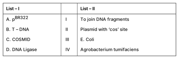
›Reveal solutionSolution
pBR322 is an E. coli plasmid, T-DNA comes from Agrobacterium tumefaciens, a cosmid is a plasmid carrying a phage cos site, and DNA ligase is the enzyme that joins DNA fragments — giving A–III, B–IV, C–II, D–I, i.e. option (B).
The concept first: the toolkit of genetic engineering
Recombinant DNA work needs three kinds of tool, and this question samples all three.
1. Vectors (the vehicles).
- pBR322 — the first widely used artificial cloning vector, built from plasmid DNA of Escherichia coli. It carries an ori, two selectable antibiotic-resistance genes (ampR and tetR) and unique restriction sites (PstI in ampR, BamHI and SalI in tetR) that allow insertional inactivation as a screen.
- Cosmid — a hybrid of a plasmid and phage λ. It is simply a plasmid that has been given the λ 'cos' (cohesive-end) site. The cos site lets the recombinant DNA be packaged inside a phage coat, so a cosmid can carry much larger inserts than an ordinary plasmid while still replicating as a plasmid inside the host.
- Ti plasmid / T-DNA — Agrobacterium tumefaciens is a soil bacterium that naturally infects dicots and transfers a piece of its tumour-inducing (Ti) plasmid — the T-DNA — into the plant genome, causing crown gall. Genetic engineers disarm the tumour genes and use the T-DNA as the delivery vehicle for a gene of interest. So T-DNA's home is Agrobacterium.
2. Enzymes (the tools).
- Restriction endonucleases are the "molecular scissors" — they cut. …
- TG EAPCET 2024Set ap-2024-05-08-FN1 markMCQQ.A person is with short stature, small round head, furrowed tongue and partially open mouth and retarded physical, psychomotor and mental development. The person is said to be suffering from this genetic disorder (A) Turner syndrome (B) Down syndrome (C) Edward syndrome (D) Klinefelter syndrome
›Reveal solutionSolution
The described symptoms — short stature, small round head, furrowed tongue, partially open mouth, and developmental delays — are classic features of Down syndrome, caused by trisomy of chromosome 21.
The question presents a clinical picture: short stature, a small round head, a furrowed tongue, a partially open mouth, and retardation in physical, psychomotor, and mental development. These are not random; they point to a specific chromosomal abnormality that affects multiple body systems. The key is to match the pattern of features, not just one symptom.
Down syndrome (trisomy 21) is the most common autosomal aneuploidy compatible with life. The extra copy of chromosome 21 disrupts development in a characteristic way. The furrowed tongue (often called a "scrotal tongue" or fissured tongue) and the partially open mouth (due to a protruding tongue and hypotonia) are particularly distinctive. The small, round head (brachycephaly) and the global developmental delay are also hallmarks.
Let's check the other options briefly to see why they don't fit:
-
Turner syndrome (45,X) affects only females. Key features are short stature, webbed neck, and lack of secondary sexual characteristics — but no furrowed tongue, small round head, or significant intellectual disability (most have normal intelligence). So it's ruled out.
-
Edward syndrome (trisomy 18) is severe and usually fatal in infancy. Features include a small head (microcephaly), low-set ears, and clenched fists with overlapping fingers. A furrowed tongue and the specific facial appearance described here are not typical. The survival is very poor, unlike the relatively longer survival in Down syndrome.
-
Klinefelter syndrome (47,XXY) affects males. Features are tall stature, small testes, gynecomastia, and often mild learning difficulties — but not short stature, small round head, or a furrowed tongue. The facial features are different. …
-
- TG EAPCET 2023Set ap-2023-05-10-FN1 markMCQQ.Match the following List – I A Glutamic acid B Lysine C Valine D Tyrosine List – II I Aromatic amino acid II Neutral amino acid III Basic amino acid IV Acidic amino acid (A) A – II, B – I, C – IV, D – III (B) A – IV, B – III, C – II, D – I (C) A – IV, B – III, C – I, D – II (D) A – I, B – IV, C – II, D – III
›Reveal solutionSolution
Amino acids are classified by the chemical nature of their side chain (R-group). Glutamic acid is acidic (IV), Lysine is basic (III), Valine is neutral (II), and Tyrosine is aromatic (I). The correct match is option (B).
The key to classifying amino acids lies in the side chain — the part that differs from one amino acid to another. The backbone (amino group, carboxyl group, and hydrogen) is identical for all standard amino acids. So the properties of each amino acid — whether it is acidic, basic, neutral, or aromatic — are entirely determined by what that R-group is.
Let’s go through each one.
-
Glutamic acid (A)
Its side chain contains a second carboxyl group (−CH2CH2COOH). At physiological pH, this extra carboxyl group loses a proton, giving the molecule a net negative charge. That makes it an acidic amino acid. So A matches with IV.
-
Lysine (B)
Its side chain is a long chain ending in an amino group (−(CH2)4NH2). This amino group can accept a proton, giving the molecule a net positive charge at physiological pH. That makes it a basic amino acid. So B matches with III.
-
Valine (C)
Its side chain is a branched hydrocarbon (−CH(CH3)2) — no charge, no aromatic ring, just a simple alkyl group. It is neither acidic nor basic, and not aromatic. That makes it a neutral (or nonpolar) amino acid. So C matches with II.
-
Tyrosine (D) …
-
- TG EAPCET 2023Set ap-2023-05-10-FN1 markMCQQ.Match the following Table - I Table - II A AUG I Phenyl alanine B UAA II Methionine C UUU III Tryptophan D UGG IV End codon (A) A – III, B – II, C – I, D – IV (B) A – II, B – III, C – IV, D – I (C) A – II, B – IV, C – I, D – III (D) A – II, B – IV, C – III, D – I
›Reveal solutionSolution
This question tests knowledge of the genetic code, specifically matching mRNA codons to their corresponding amino acids or functions. The correct match is AUG to Methionine, UAA to End codon, UUU to Phenylalanine, and UGG to Tryptophan, leading to option (C).
Concept and Intuition
The genetic code is a set of rules by which information encoded in genetic material (DNA or RNA sequences) is translated into proteins by living cells. This information is carried in sequences of three nucleotides called codons. Each codon typically specifies a particular amino acid, or signals the start or termination of protein synthesis.
Understanding the genetic code is fundamental to molecular biology. Key aspects include:
- Triplet nature: Each codon consists of three nucleotides.
- Specificity: Each codon codes for only one specific amino acid (or a stop signal).
- Degeneracy: Most amino acids are coded by more than one codon.
- Universality: The genetic code is largely the same across all forms of life.
- Start codon: AUG typically initiates translation and codes for Methionine.
- Stop codons: UAA, UAG, and UGA signal the termination of translation.
To solve this problem, we need to recall the specific amino acids or functions associated with the given mRNA codons.
Step-by-step Matching
-
Analyze Codon A: AUG
- AUG is universally recognized as the start codon in mRNA.
- It also codes for the amino acid Methionine (Met).
- Therefore, A matches with II (Methionine).
-
Analyze Codon B: UAA
- UAA is one of the three stop codons (the others being UAG and UGA).
- Stop codons do not code for any amino acid; instead, they signal the termination of protein synthesis. They are also referred to as "end codons" or "nonsense codons".
- Therefore, B matches with IV (End codon).
-
Analyze Codon C: UUU
- UUU is a codon that codes for the amino acid Phenylalanine (Phe). …
- TG EAPCET 2023Set ap-2023-05-10-AN1 markMCQQ.Select the correct statements from the following I) RNA also functions as adapter molecule II) Base pairing confers unique property to the polynucleotide chains III) DNA replication occurs after chromosomal division IV) DNA replication occurs frequently in a cell (A) II & IV (B) I & II (C) II & III (D) I & III
›Reveal solutionSolution
The key idea is to evaluate each statement against established molecular biology facts. Only statements I and II are correct, so the answer is option (B).
Let’s break down each statement one by one, with the underlying concept first.
Concept: Adapter molecules in translation
In protein synthesis, tRNA (transfer RNA) acts as an "adapter" — it carries a specific amino acid at one end and has an anticodon at the other that base-pairs with the mRNA codon. This is the classic adapter function. But the statement says "RNA also functions as adapter molecule." The word "also" is key: it doesn't say only tRNA; it says RNA also does this. And indeed, besides tRNA, other RNAs like rRNA (in ribosomes) and even some small RNAs have adapter-like roles in specific contexts. However, the most direct and widely taught adapter is tRNA. Statement I is correct.
Concept: Base pairing and polynucleotide structure
Base pairing (A with T/U, G with C) is what gives DNA and RNA their double-stranded stability and specificity. It allows complementary strands to form, enables replication, transcription, and the genetic code. Without base pairing, polynucleotide chains would just be random polymers. So statement II is correct.
Concept: Timing of DNA replication relative to cell division
DNA replication occurs before chromosomal division (mitosis or meiosis), not after. During the S phase of the cell cycle, the entire genome is duplicated. Then, in M phase, the duplicated chromosomes separate. So statement III is false — replication precedes division, not the other way around.
Concept: Frequency of DNA replication in a cell …
- TG EAPCET 2023Set ap-2023-05-11-AN1 markMCQQ.In a transcription unit the coding strand is A. Found in DNA B. Found in RNA C. Codes for hnRNA D. Does not code for anything Which of the above statements are true (A) AC (B) AB (C) AD (D) BD
›Reveal solutionSolution
The coding strand is one of the two DNA strands in a transcription unit and is not directly transcribed by RNA polymerase. Therefore, statements A ("Found in DNA") and D ("Does not code for anything" in the sense of being transcribed) are true. The correct option is (C).
Concept and Intuition
A transcription unit in DNA is a segment that is transcribed into an RNA molecule. It typically consists of three main regions:
- Promoter: The binding site for RNA polymerase, located upstream of the structural gene.
- Structural gene: The region that actually codes for the RNA molecule.
- Terminator: The region that signals the end of transcription.
Within the structural gene, the DNA is double-stranded. During transcription, only one of these strands serves as a template for RNA synthesis. These two strands are given specific names:
- Template strand (or antisense strand): This is the DNA strand that is actually read by RNA polymerase. Its sequence is complementary to the RNA molecule being synthesized. RNA polymerase moves along this strand in the 3′→5′ direction, synthesizing RNA in the 5′→3′ direction.
- Coding strand (or sense strand): This is the DNA strand that is not transcribed. Its sequence is identical to the RNA molecule being synthesized, with the only difference being that DNA has thymine (T) where RNA has uracil (U). It's called the "coding" strand because its sequence directly represents the codons that will be found in the mRNA and translated into protein. However, it is not the strand that RNA polymerase uses as a template.
The product of transcription in eukaryotes is initially heterogeneous nuclear RNA (hnRNA), which then undergoes processing (splicing, capping, tailing) to become mature messenger RNA (mRNA).
Step-by-step Evaluation of Statements
Let's evaluate each statement regarding the coding strand:
-
Statement A: Found in DNA.
- The coding strand is one of the two strands that make up the DNA double helix within a transcription unit. It is a DNA strand.
- Conclusion: Statement A is True.
-
Statement B: Found in RNA.
- The coding strand is a DNA strand. RNA is a separate molecule synthesized from the template DNA strand. The coding strand itself is not RNA.
- Conclusion: Statement B is False.
-
Statement C: Codes for hnRNA. …
🎓Unlock everything free for 14 days
- ✓Full step-by-step solutions
- ✓Concept-first explanations
- ✓Methods, shortcuts & mistakes
- ✓PYQ mapping + timed mock tests
Full access for 14 days. No credit card required.