Q.Given below is the sequence of coding strand of DNA in a transcription unit: 3'-AATGCAGCTATTAGG-5'. Write the sequence of
🔒You're viewing a preview — the full solution, concept, methods & PYQ mapping are locked.
🔒 Start your 14-day free trial to unlock the full solution →Concept understanding — Template Strand Transcription
Template Strand Transcription: A First Look
Imagine you have a master recipe book written in a language only the head chef can read. To share a recipe with the kitchen staff, you don't hand them the original book — you make a working copy on a separate sheet, using the original as your guide. That original page you read from is the template. The copy you produce is the transcript.
In a cell, the master recipe book is DNA. It holds all the instructions for making proteins, which do almost everything in your body. But DNA never leaves the nucleus — it's too precious and too large. So the cell makes a temporary, portable copy of a specific instruction. That copy is called messenger RNA (mRNA).
The process of making this mRNA copy is transcription. And the strand of DNA that is actually read to make the copy is called the template strand.
The Precise Meaning
DNA is a double helix — two strands twisted together. During transcription, the cell unzips a small section of this helix. Only one of the two strands serves as the blueprint. That strand is the template strand. The other strand, called the coding strand (or non-template strand), is not read — it just sits there, matching the sequence of the mRNA that gets made (with one chemical difference: DNA uses T, RNA uses U).
So the template strand is the actual DNA sequence that RNA polymerase (the enzyme that does the copying) reads and uses to build a complementary mRNA molecule.
Think of the template strand as the negative of a photograph. The mRNA is the print made from that negative. The coding strand is like a second print that happens to look almost identical to the final photo — but it wasn't used to make it.
Why It Matters
- Accuracy: The cell must read the correct strand. If it read the wrong one, the mRNA would be nonsense and the protein would be wrong or non-functional.
- Direction: RNA polymerase reads the template strand in the 3' to 5' direction, and builds mRNA in the 5' to 3' direction. This is a fixed rule — like reading a sentence left to right.
- Gene regulation: Which strand is the template for a given gene is fixed. But different genes on the same DNA molecule may use different strands as their template. So a single stretch of DNA can contain genes pointing in opposite directions.
A Simple Example
Suppose a short stretch of DNA has these two strands:
- Strand A:
ATGCGT - Strand B:
TACGCA
If Strand A is the template, the mRNA made will be complementary to it: UACGCA (remember, U replaces T in RNA).
If Strand B is the template, the mRNA will be complementary to B: AUGCGU.
The two mRNAs are completely different. So the cell must know, for each gene, which strand is the template. That information is encoded in the DNA sequence itself — in the promoter region that tells RNA polymerase where to start and which way to go.
The template strand is not the same as the coding strand. The mRNA sequence is identical to the coding strand (with U instead of T), but it is complementary to the template strand. This is a common confusion — the mRNA looks like the coding strand, but it was built from the template strand.
What NCERT Says …
First, recall the key distinction: the coding strand (also called the sense strand) has the same sequence as the mRNA (except T is replaced by U in RNA). The template strand is the one actually used by RNA polymerase to synthesise mRNA — it is complementary and antiparallel to the coding strand.
- The given coding strand is written 3'-AATGCAGCTATTAGG-5'. Pairing each base directly opposite it (A-T, G-C) at the same left-to-right position gives the template strand, read 5' to 3': 5'-TTACGTCGATAATCC-3'.
- For the mRNA, first rewrite the coding strand in the conventional 5' to 3' direction by reversing the given sequence: 5'-GGATTATCGACGTAA-3'. The mRNA is identical to this, base for base, except thymine (T) is replaced by uracil (U). …
In a transcription unit, the coding strand of DNA has the same sequence as the mRNA (except T replaced by U), while the complementary strand serves as the template for RNA synthesis.
When we talk about transcription, we are describing the process by which a segment of DNA is copied into RNA — specifically, messenger RNA (mRNA), in the case of protein-coding genes. The DNA in a transcription unit is double-stranded, but only one of the two strands acts as the template for RNA synthesis; the other, the coding (sense) strand, is not copied but has the same sequence as the resulting mRNA (except thymine is replaced by uracil).
The sequence given, 3'-AATGCAGCTATTAGG-5', is stated to be the coding strand.
(a) The complementary (template) strand. DNA's two strands are antiparallel and complementary: wherever one strand reads A, the other reads T at the same physical position, and wherever one reads G, the other reads C. Pairing every base of the given strand this way, at the same left-to-right position, gives the complementary strand — but since the strands run in opposite directions, this paired strand must be labelled 5' at the end where the coding strand is labelled 3', and vice versa:
3'-A A T G C A G C T A T T A G G-5' (coding strand, given)
5'-T T A C G T C G A T A A T C C-3' (complementary/template strand)
So the complementary strand is 5'-TTACGTCGATAATCC-3'.
A common mistake here is to write the complementary strand in the same 3' to 5' orientation as the given strand. Because the two strands of DNA are antiparallel, the complementary strand's 5' end sits opposite the coding strand's 3' end. …
Work directly from the mRNA-equals-coding-strand rule instead of computing the template strand first: keep the same left-to-right letter order as the given coding strand and replace each base with its Watson-Crick partner (A<->T, G<->C) -- only the end-labels flip, not the letter order -- to get the template …
- TG EAPCET 2025Set ap-2025-04-29-AN1 markMCQQ.Identify the wrong statements from the following I. DNA polymerase has capability of catalysing the process of elongation of polypeptide chain II. In splicing, introns are removed and exons are joined III. In capping, methyl guanosine triphosphate is added to the 5' end of hnRNA IV. In tailing, adenylate residues are added at 5' end (A) I & II (B) II & III (C) I & IV (D) III & IV
›Reveal solutionSolution
The key idea is to recall the specific roles of DNA polymerase and the three post-transcriptional modifications in eukaryotes. DNA polymerase works on DNA, not polypeptide chains; tailing adds adenylate residues to the 3' end, not the 5' end. So statements I and IV are wrong.
Let’s go through each statement one by one, checking it against what actually happens in molecular biology.
-
Statement I: “DNA polymerase has capability of catalysing the process of elongation of polypeptide chain”
This is false. DNA polymerase is an enzyme that synthesizes DNA — it adds deoxyribonucleotides to a growing DNA strand during replication or repair. The elongation of a polypeptide chain (protein synthesis) is catalysed by the ribosome (specifically, the peptidyl transferase activity of the rRNA in the large subunit). DNA polymerase has nothing to do with proteins. So I is wrong.
-
Statement II: “In splicing, introns are removed and exons are joined”
This is correct. In eukaryotic gene expression, the primary transcript (pre-mRNA or hnRNA) contains both introns (non-coding) and exons (coding). Splicing is the process where introns are cut out and exons are ligated together to form the mature mRNA. So II is right.
-
Statement III: “In capping, methyl guanosine triphosphate is added to the 5' end of hnRNA” …
-
- TG EAPCET 2025Set ap-2025-04-29-FN1 markMCQQ.Select the correct pair (A) Genetic nature of DNA → Frankel-Conrat (B) In vitro DNA synthesis → Hanning (C) Double helical model of RNA → Watson and Crick (D) Genetic code → Marshall Nirenberg
›Reveal solutionSolution
The question asks to identify the correct pairing of a scientific discovery with its discoverer. The correct pair is the genetic code associated with Marshall Nirenberg.
Understanding the history of major discoveries in molecular biology, along with the scientists who made them, is fundamental. Many key concepts in genetics and molecular biology are named after or strongly associated with specific researchers and their groundbreaking experiments. This question tests your knowledge of these historical milestones.
Let's evaluate each option:
-
Option (A): Genetic nature of DNA → Frankel-Conrat
- The "genetic nature of DNA" refers to the discovery that DNA is the hereditary material responsible for transmitting genetic information from one generation to the next. This was established through a series of experiments:
- Frederick Griffith (1928): Demonstrated transformation in bacteria, suggesting a "transforming principle."
- Avery, MacLeod, and McCarty (1944): Identified DNA as the transforming principle.
- Hershey and Chase (1952): Provided definitive proof that DNA, not protein, is the genetic material in bacteriophages.
- Heinz Fraenkel-Conrat (along with B. Singer) conducted experiments in 1956 with the Tobacco Mosaic Virus (TMV). They showed that in TMV, RNA, not DNA, is the genetic material. They separated the RNA and protein components of the virus, recombined them, and demonstrated that the type of progeny virus produced was determined by the RNA, not the protein coat.
- Therefore, Frankel-Conrat's work established RNA as the genetic material in some viruses, not the general genetic nature of DNA. This option is incorrect.
- The "genetic nature of DNA" refers to the discovery that DNA is the hereditary material responsible for transmitting genetic information from one generation to the next. This was established through a series of experiments:
-
Option (B): In vitro DNA synthesis → Hanning
- "In vitro DNA synthesis" refers to the artificial synthesis of DNA in a test tube, outside of a living organism.
- Arthur Kornberg was awarded the Nobel Prize in 1959 for his discovery of the mechanisms in the biological synthesis of deoxyribonucleic acid. He isolated and characterized DNA polymerase I, the enzyme responsible for synthesizing new DNA strands from a DNA template and nucleotide precursors in vitro.
- Elisabeth Hanning (1904) is known for her pioneering work in plant tissue culture, specifically the in vitro culture of immature plant embryos (embryo culture). Her work was foundational for plant biotechnology.
- Therefore, Hanning is not associated with in vitro DNA synthesis. This option is incorrect.
-
Option (C): Double helical model of RNA → Watson and Crick
- James Watson and Francis Crick are famously known for proposing the double helical model of DNA in 1953, based on X-ray diffraction data from Rosalind Franklin and Maurice Wilkins, and chemical data from Erwin Chargaff. …
-
- TG EAPCET 2025Set ap-2025-04-29-FN1 markMCQQ.The recombination frequency between the genes a and c is 5%; b and c is 15%; b and d is 9%; a and b is 20%; c and d is 24%; and a and d is 29%; Then identify the sequence of genes on a linear chromosome? (A) a, d, b, c (B) d, b, a, c (C) a, c, b, d (D) a, b, c, d
›Reveal solutionSolution
The key idea is that recombination frequency is proportional to physical distance, so the smallest frequencies indicate the closest gene pairs. By placing the closest pairs together and checking consistency, the correct gene order is a, c, b, d — option (C).
The concept here is gene mapping on a linear chromosome. Recombination frequency (RF) between two genes tells you how far apart they are — the higher the RF, the farther apart they are. On a linear chromosome, distances are additive: if you know the distances between all pairs, you can deduce the order by finding which arrangement makes the distances consistent.
We have six RF values:
- a–c: 5%
- b–c: 15%
- b–d: 9%
- a–b: 20%
- c–d: 24%
- a–d: 29%
The smallest RF is 5% between a and c, so a and c are the closest pair. That means they must be neighbours on the chromosome. Let’s place them as a–c or c–a for now.
Next, look at b. b is 15% from c and 20% from a. If a and c are 5 apart, then b could be on either side of the pair. If b is on the a side, then distance a–b should be small, but it’s 20 — that’s larger than a–c (5). If b is on the c side, then b–c should be smaller than a–b, which matches: b–c = 15, a–b = 20. So b is on the c side: order a–c–b.
Now check distances: a–c = 5, c–b = 15, so a–b should be 5 + 15 = 20 — exactly matches the given 20%. Good. …
- TG EAPCET 2025Set ap-2025-04-30-FN1 markMCQQ.Match the list-1 with list-2 and select the correct option List-1 List-2 A. Operator site I. Binding site for RNA polymerase B. Promotor site II. Binding site for repressor molecule C. Regulator gene III. Codes for protein/Enzyme D. Structural gene IV. Codes for repressor molecule (A) A – II, B – I, C – IV, D – III (B) A – II, B – I, C – III, D – IV (C) A – I, B – IV, C – II, D – III (D) A – III, B – II, C – I, D – IV
›Reveal solutionSolution
The lac operon model defines four key genetic elements: the operator binds the repressor, the promoter binds RNA polymerase, the regulator gene codes for the repressor, and structural genes code for enzymes. The correct match is (A).
Gene regulation in prokaryotes, famously illustrated by the lac operon in E. coli, relies on a coordinated set of DNA sequences and genes working together. Understanding what each component does makes matching straightforward.
The promoter is where transcription begins. RNA polymerase recognizes and binds to this site to initiate mRNA synthesis. Without a functional promoter, the downstream genes remain silent.
The operator sits adjacent to the promoter and acts as a molecular switch. When a repressor protein binds here, it physically blocks RNA polymerase from transcribing the structural genes. Think of it as a gate that can be closed by the repressor.
The regulator gene (often denoted lacI in the lac operon) is located upstream and codes for the repressor protein itself. This gene is transcribed independently and produces the molecule that can bind to the operator. …
- TG EAPCET 2023Set ap-2023-05-10-FN1 markMCQQ.Below Diagram shows important concept of genetic implication of DNA. Fill in the blanks A, B, C [FIGURE] (A) Transcription → (B) Translation → (C) Crick (B) Translation → (B) Extension → (C) Franklin (C) Transcription → (B) Replication → (C) Watson (D) Translation → (B) Transcription → (C) Chargaff
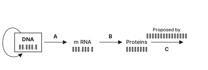
›Reveal solutionSolution
The diagram illustrates the central dogma of molecular biology (DNA → RNA → Protein), so the blanks must be filled as Transcription, Translation, and Crick — the scientist who proposed this flow. The correct option is (A).
The question asks you to identify the correct sequence of processes and the scientist associated with the central dogma of molecular biology. The diagram likely shows a flow from DNA to a functional product, with blanks for the two key processes and the name of the scientist who first articulated this concept.
Concept & Intuition:
The central dogma, famously stated by Francis Crick, describes the directional flow of genetic information: DNA is transcribed into RNA, which is then translated into protein. This is the fundamental framework for gene expression. The other options mix up processes (e.g., replication, extension) or attribute the dogma to the wrong scientist (Franklin, Watson, Chargaff — each famous for other contributions: Franklin for X-ray crystallography of DNA, Watson for the double-helix model, Chargaff for base-pairing rules).
Step-by-step reasoning:
-
Identify the first blank (A): The process that converts DNA into RNA is transcription. This rules out options that start with "Translation" (B and D), because translation uses RNA to make protein, not DNA to RNA.
-
Identify the second blank (B): After transcription, the RNA molecule is used to direct protein synthesis via translation. So the second process must be translation. This eliminates option (C), which has "Replication" (copying DNA) in the second blank. …
-
- TG EAPCET 2022Set ap-2022-07-30-FN1 markMCQQ.Match the following : List-I A) Free phosphate moiety polymer B) Free OH group end C) The right handed fashion coiling D) The flow of information of gene in Retroviruses List-II I) From RNA to DNA II) 5′ end of the polynucleotide chain III) 3′ end of the polynucleotide chain IV) Helix has 3.4 nm pitch The correct match is: (A) I B) IV C) II D) III (B) IV B) III C) II D) I (C) II B) IV C) III D) I (D) II B) III C) IV D) I
›Reveal solutionSolution
This question matches molecular biology terms (free phosphate, free OH, right-handed helix, retrovirus gene flow) with their correct descriptions. The correct pairing is A→II, B→III, C→IV, D→I, which corresponds to option (D).
Concept & Intuition
The problem tests basic knowledge of nucleic acid structure and the central dogma in retroviruses.
- A polynucleotide chain has a 5′ end (free phosphate) and a 3′ end (free hydroxyl).
- The classic DNA double helix is right-handed with a pitch of 3.4 nm (one full turn per 10 base pairs).
- Retroviruses (like HIV) reverse the usual flow: they use reverse transcriptase to go from RNA to DNA, not DNA to RNA.
Step-by-step reasoning
-
A) Free phosphate moiety polymer
- In a polynucleotide, the phosphate group is attached to the 5′ carbon of the sugar. The end with a free phosphate is the 5′ end.
- So A matches II (5′ end of the polynucleotide chain).
-
B) Free OH group end
- The other end of the chain has a free hydroxyl (–OH) on the 3′ carbon of the sugar. This is the 3′ end.
- So B matches III (3′ end of the polynucleotide chain).
-
C) The right handed fashion coiling
- The B‑form DNA helix is right‑handed and has a pitch of 3.4 nm (the distance for one complete turn).
- So C matches IV (Helix has 3.4 nm pitch).
-
D) The flow of information of gene in Retroviruses …
- TG EAPCET 2022Set ap-2022-07-31-FN1 markMCQQ.Assertion (A): In DNA double helix, one full turn of helical strand contains twelve base pairs. Reason (R): The pitch of DNA helix is 34∘ A. The correct option among the following is: (A) is true, (R) is true and (R) is the correct explanation for (A) (B) (A) is true, (R) is true but (R) is not the correct explanation for (A) (C) (A) is true but (R) is false (D) (A) is false but (R) is true
›Reveal solutionSolution
The assertion that one full turn of the DNA double helix contains twelve base pairs is false (it actually contains 10 base pairs), and the reason stating the pitch is 34 Å is true but irrelevant to the false assertion. Therefore, the correct option is (D).
The key here is to recall the classic B‑form DNA structure, which is the most common form under physiological conditions. The double helix has a pitch (the length of one complete turn) of 34 Å, and the rise per base pair is 3.4 Å. Dividing the pitch by the rise gives the number of base pairs per turn:
3.4 A˚/base pair34 A˚=10 base pairs.
So one full turn contains 10 base pairs, not 12. The assertion (A) is therefore false.
The reason (R) states that the pitch is 34 Å — this is a well‑established fact for B‑DNA, so (R) is true. However, even if (R) were correct, it does not explain a false statement.
Let’s go step by step:
-
Check the Assertion (A):
“In DNA double helix, one full turn of helical strand contains twelve base pairs.”
- In B‑DNA, the number of base pairs per turn is 10. In A‑DNA it is about 11, and in Z‑DNA it is 12, but the question refers to the standard double helix (B‑form).
- Therefore, (A) is false.
-
Check the Reason (R):
“The pitch of DNA helix is 34 Å.”
- The pitch of B‑DNA is indeed 34 Å (the distance along the helix axis for one complete turn).
- So (R) is true.
-
Determine the relationship: …
-
- TG EAPCET 2022Set ap-2022-07-31-AN1 markMCQQ.The DNA molecule has 4 heterocyclic compounds namely ‘A’, ‘B’, ‘C’, and ‘D’. In that ‘A’ pairs with ‘C’ by means of 2 hydrogen bonds, while ‘B’ pairs with ‘D’ by means of 3 hydrogen bonds during formation of its double helical structure. Identify the names of ‘A’, ‘B’, ‘C’, and ‘D’, in the same sequence. (A) Adenine, Guanine, Thymine and Cytosine (B) Guanine, Adenine, Cytosine and Thymine (C) Thymine, Adenine, Guanine and Cytosine (D) Cytosine, Guanine, Adenine and Thymine
›Reveal solutionSolution
DNA base pairing follows Chargaff's rules: purines pair with pyrimidines via specific hydrogen bonds. Adenine pairs with Thymine (2 H-bonds) and Guanine pairs with Cytosine (3 H-bonds). The answer is (A).
The structure of DNA relies on complementary base pairing between nitrogenous bases on opposite strands. Two purines (adenine and guanine, with double-ring structures) pair with two pyrimidines (thymine and cytosine, with single-ring structures). The number of hydrogen bonds between paired bases is fixed by their chemical structure: smaller bases form fewer bonds, larger complementary pairs form more.
The key is recognizing which base pairs with which, and how many hydrogen bonds stabilize each pair.
-
Identify the pairing pattern from hydrogen bond count.
The problem states that A pairs with C via 2 hydrogen bonds, and B pairs with D via 3 hydrogen bonds. In DNA, only two pairing combinations exist:
- Adenine (A) ↔ Thymine (T): 2 hydrogen bonds
- Guanine (G) ↔ Cytosine (C): 3 hydrogen bonds
-
Match the 2-hydrogen-bond pair.
Since 'A' pairs with 'C' through 2 hydrogen bonds, this must be the adenine–thymine pair. Therefore:
- 'A' = Adenine
- 'C' = Thymine
-
Match the 3-hydrogen-bond pair. …
-
- TG EAPCET 2021Set ap-2021-08-09-FN1 markMCQQ.In a striated myofibril, the F-actin of thin filament is formed by (A) G-actin (B) Meromyosin (C) Troponin-C (D) Tn-I
›Reveal solutionSolution
The thin filament in a striated myofibril is built from G-actin monomers that polymerize into F-actin (filamentous actin). The correct option is (A).
The question asks what forms the F-actin of the thin filament. This is a classic structure-function question from muscle biology. The thin filament is a complex assembly, but its backbone is a double helix of actin. That actin exists in two forms: globular actin (G-actin), the monomer, and filamentous actin (F-actin), the polymer. The F-actin strand is literally made by G-actin subunits joining end-to-end.
Let’s walk through the options to see why only one fits.
-
G-actin is the monomeric form of actin. In muscle cells, these globular subunits polymerize to form the long, helical F-actin filament. So F-actin is formed by G-actin — this is the direct answer.
TipThink of G-actin as bricks and F-actin as the wall. The wall is made of bricks, not the other way around.
-
Meromyosin is a fragment of the myosin heavy chain (the thick filament protein). It has nothing to do with building actin filaments. It’s involved in the cross-bridge cycle, not thin filament structure. …
-
🎓Unlock everything free for 14 days
- ✓Full step-by-step solutions
- ✓Concept-first explanations
- ✓Methods, shortcuts & mistakes
- ✓PYQ mapping + timed mock tests
Full access for 14 days. No credit card required.