Q.Draw a well-labelled diagram of an antibody molecule.
You're viewing a preview — the full solution, concept, methods & PYQ mapping are locked.
Start your 14-day free trial to unlock the full solution →An antibody (immunoglobulin) is a Y-shaped protein molecule made of four polypeptide chains—two identical heavy chains and two identical light chains—joined by disulfide bonds, with variable regions at the tips that bind specific antigens and a constant region that determines effector functions.
Antibodies are the molecular soldiers of our adaptive immune system, produced by plasma cells (differentiated B lymphocytes) in response to foreign antigens. Each antibody is exquisitely specific, recognizing and binding to a unique epitope on an antigen—be it a viral protein, bacterial toxin, or any other foreign molecule. Understanding their structure reveals how they achieve this remarkable specificity while also triggering immune responses.
The basic structural unit of an antibody is a monomer composed of four polypeptide chains. Two of these are identical heavy chains (H chains), each about 450–550 amino acids long, and two are identical light chains (L chains), each about 220 amino acids long. These chains are held together by disulfide bonds (covalent S–S linkages between cysteine residues) and non-covalent interactions, forming the characteristic Y-shaped molecule.
Each chain—both heavy and light—has two distinct regions. The variable region (V region) occupies the amino-terminal end and shows tremendous diversity in amino acid sequence from one antibody to another. This variability is concentrated in three hypervariable regions within each V domain, called complementarity-determining regions (CDRs), which directly contact the antigen. The constant region (C region) makes up the rest of the chain and has a relatively uniform sequence within a given antibody class.
At the tips of the Y, where one heavy-chain variable region (VH) pairs with one light-chain variable region (VL), we find the antigen-binding sites. Each antibody monomer has two such sites, making it bivalent—it can bind two identical epitopes simultaneously. The precise three-dimensional pocket formed by the CDRs of VH and VL determines which antigen the antibody recognizes, much like a lock accepts only a specific key.
The stem of the Y is formed entirely by the constant regions of the two heavy chains. This Fc region (fragment crystallizable) does not bind antigen but instead interacts with other components of the immune system—complement proteins, phagocytic cells bearing Fc receptors, and so on. It is this region that determines the antibody's class (IgG, IgM, IgA, IgE, or IgD) and thus its effector functions: whether it can cross the placenta, activate complement efficiently, or trigger mast cell degranulation.
The two arms of the Y, each containing one complete light chain and the VH plus part of the constant region of a heavy chain, are called Fab regions (fragment antigen-binding). A flexible hinge region between the Fab and Fc portions allows the two antigen-binding sites to operate somewhat independently, accommodating antigens that may be spaced at varying distances on a pathogen's surface.
Labelled Diagram of an Antibody Molecule
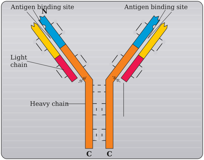
Key labels (see the figure above):
Key labels:
- Heavy chain (H): The longer polypeptide, with one variable domain (VH) and three or four constant domains (CH1, CH2, CH3, and sometimes CH4).
- Light chain (L): The shorter polypeptide, with one variable domain (VL) and one constant domain (CL). …
Unlock everything free for 14 days
- Full step-by-step solutions
- Concept-first explanations
- Methods, shortcuts & mistakes
- PYQ mapping + timed mock tests
Full access for 14 days. No credit card required.