Biology · Ch 5 — Molecular Basis of Inheritance
Types of RNA and the process of Transcription
Types of RNA and the process of Transcription
The Three Major Types of RNA in Bacteria
In a bacterial cell, three distinct kinds of RNA work together to build a protein. Messenger RNA (mRNA) provides the actual template — the sequence of codons that will be read. Transfer RNA (tRNA) brings the correct amino acids to the growing protein chain and reads the genetic code on the mRNA. Ribosomal RNA (rRNA) plays both a structural and a catalytic role inside the ribosome during translation. All three are essential; none can be skipped.
Transcription in Bacteria — One Enzyme Does It All
Bacteria have a single DNA-dependent RNA polymerase that catalyses the transcription of all three types of RNA. This enzyme uses nucleoside triphosphates as substrates and polymerises RNA in a template-dependent fashion, following the rule of complementarity (A pairs with U, T pairs with A, C pairs with G, G pairs with C).
The process has three stages:
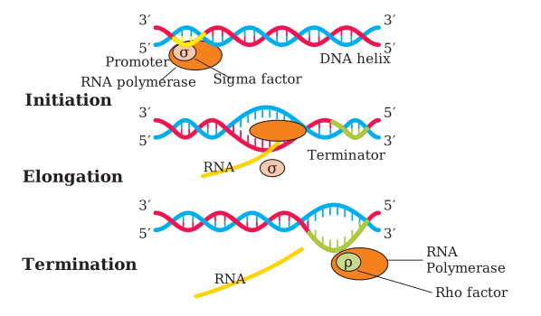
- Initiation: RNA polymerase binds to a specific DNA sequence called the promoter and begins transcription.
- Elongation: The enzyme somehow helps open the DNA helix and continues adding nucleotides. Only a short stretch of newly made RNA remains bound to the enzyme at any time.
- Termination: When the polymerase reaches a terminator region on the DNA, the nascent RNA falls off, and the RNA polymerase itself detaches. Transcription stops.
How the Same Enzyme Does Three Different Jobs
Here is an intriguing puzzle: RNA polymerase can only catalyse elongation by itself. How then does it manage initiation and termination? The answer lies in two transient helper factors.
- Initiation factor (σ, sigma): This factor associates with RNA polymerase temporarily, changing its specificity so that it recognises the promoter and starts transcription.
- Termination factor (ρ, rho): This factor associates with the polymerase later, altering its specificity so that it recognises the terminator and stops transcription.
Without these factors, the core enzyme can only elongate — it cannot start or stop on its own.
Coupled Transcription and Translation in Bacteria
Because bacteria have no nucleus, transcription and translation occur in the same compartment (the cytosol). Moreover, bacterial mRNA does not require any processing to become active. As a result, translation can begin on the mRNA even before the full mRNA molecule has been transcribed. This means transcription and translation are coupled in bacteria — a ribosome can start reading the 5' end of the mRNA while the 3' end is still being synthesised.
The Extra Complexities in Eukaryotes
Eukaryotic cells face two major additional complexities that bacteria do not.
First complexity — multiple RNA polymerases with division of labour. The nucleus contains at least three distinct RNA polymerases (organelles like mitochondria have their own separate RNA polymerase).
- RNA polymerase I transcribes the genes for ribosomal RNAs (28S, 18S, and 5.8S rRNAs).
- RNA polymerase II transcribes the precursor of mRNA, which is called heterogeneous nuclear RNA (hnRNA).
- RNA polymerase III transcribes tRNA, 5S rRNA, and small nuclear RNAs (snRNAs).
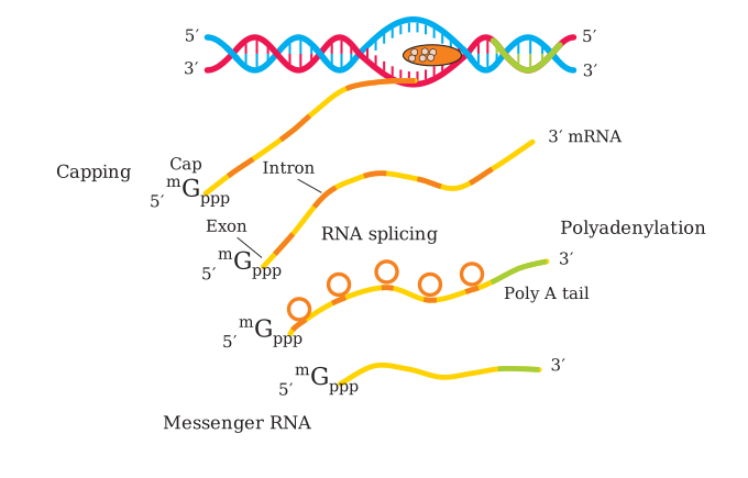
Second complexity — the primary transcript must be processed. The initial RNA transcript (hnRNA) contains both exons (coding sequences) and introns (non-coding intervening sequences). This primary transcript is non-functional. It must undergo three processing steps before it becomes mature mRNA.
- Splicing: The introns are removed, and the exons are joined together in a defined order.
- Capping: An unusual nucleotide — methyl guanosine triphosphate — is added to the 5' end of the hnRNA.
- Tailing: Adenylate residues (200–300 of them) are added to the 3' end in a template-independent manner.
Only after this full processing is the hnRNA called mRNA. It is then transported out of the nucleus into the cytoplasm for translation.
Why These Complexities Matter
The split-gene arrangement (exons interrupted by introns) is probably an ancient feature of the genome. The presence of introns is reminiscent of antiquity, and the process of splicing represents the dominance of an RNA world in early evolution. In recent times, the understanding of RNA and RNA-dependent processes in living systems has assumed much greater importance.
The Genetic Code — A Triplet Code
During replication and transcription, one nucleic acid is copied to form another nucleic acid, so complementarity makes these processes easy to understand. But translation requires transferring information from a polymer of nucleotides to a polymer of amino acids. No complementarity exists between nucleotides and amino acids, nor could any be drawn theoretically. Yet evidence strongly supported the idea that changes in nucleic acids (genetic material) caused changes in amino acids in proteins. This led to the proposition of a genetic code that could direct the sequence of amino acids during protein synthesis.
The Triplet Hypothesis
It was George Gamow, a physicist, who argued that since there are only four bases and they must code for 20 amino acids, the code must be a combination of bases. He proposed that the code should be made up of three nucleotides — a triplet. This was a bold proposition because a permutation combination of four cubed (4 × 4 × 4) would generate 64 codons — many more than the 20 needed.
Deciphering the Code
Providing proof that the codon was a triplet was a daunting task. The chemical method developed by Har Gobind Khorana was instrumental in synthesising RNA molecules with defined combinations of bases (homopolymers and copolymers). Marshall Nirenberg's cell-free system for protein synthesis finally helped decipher the code. Severo Ochoa's enzyme (polynucleotide phosphorylase) was also helpful in polymerising RNA with defined sequences in a template-independent manner (enzymatic synthesis of RNA). The final result was a checkerboard for the genetic code.
Salient Features of the Genetic Code
- The codon is triplet. 61 codons code for amino acids, and 3 codons do not code for any amino acid — they function as stop codons.
- The code is degenerate. Some amino acids are coded by more than one codon.
- The code is read in a contiguous fashion. There are no punctuations between codons. …
Drawn by us to help you understand the concept clearly, and verified to make sure it's accurate. For exams, practice from your textbook's own diagram.
Figure 5.10 is a three-panel diagram (labelled, left to right, Initiation, Elongation, and Termination) that walks through bacterial transcription as a single continuous process on one DNA molecule.
In the Initiation panel, the DNA double helix is still fully formed. RNA polymerase is shown bound at the promoter together with a small attached shape representing the sigma factor (σ) — the transient partner that lets the core enzyme recognise the promoter and start transcription.
In the Elongation panel, the helix is shown locally opened around the polymerase, and a short stretch of newly made RNA (drawn as a curling strand) emerges from the enzyme as it moves toward the terminator. The sigma factor is shown separately/released at this stage, since it is no longer needed once transcription is under way.
In the Termination panel, the opened region of the helix is larger, a longer RNA strand trails from the complex, and a second small attached shape — the rho factor (ρ) — is shown together with the polymerase at the terminator, marking the point where the enzyme releases the finished RNA and detaches from the DNA.
The figure's teaching point is narrow and precise: RNA polymerase can only catalyse elongation on its own; sigma and rho are the two transient helper factors that let the same enzyme also start (initiation) and stop (termination) transcription. …
Drawn by us to help you understand the concept clearly, and verified to make sure it's accurate. For exams, practice from your textbook's own diagram.
The figure shows a schematic of the nucleus of a eukaryotic cell, with the nuclear envelope drawn as a boundary. Inside the nucleus, a DNA double helix is depicted with its two strands. One strand serves as the template for transcription. An RNA polymerase II enzyme is shown bound to the DNA at a promoter region, actively synthesising a long primary transcript called hnRNA (heterogeneous nuclear RNA). This hnRNA is drawn as a continuous wavy line emerging from the polymerase, and it contains both exons (coding segments, often shown as thicker or coloured blocks) and introns (non-coding segments, shown as thinner or differently coloured stretches interspersed between exons).
The figure then illustrates three processing steps that happen to the hnRNA while it is still inside the nucleus:
- Capping – A small labelled structure (methyl guanosine triphosphate) is added to the 5'-end of the hnRNA.
- Tailing – A string of adenylate residues (200–300 As) is added to the 3'-end in a template-independent manner.
- Splicing – The introns are removed, and the exons are joined together in a precise order. This is often shown by arrows or a separate panel where the introns loop out and are cut away, leaving only the exons connected.
After these three modifications, the hnRNA has become mature mRNA. The figure then shows this mature mRNA exiting the nucleus through a nuclear pore (often drawn as a gap or channel in the nuclear envelope) into the cytoplasm, where it will be translated into protein. …