Question of 96
Q.Draw a neat labelled diagram of DNA double Helix.
Goa GbshseGBSHSE Class 12 Board Exam 2024Subjective· 2mImportance★★★★★
0% · 0/96 Questions
You're viewing a preview — the full solution, concept, methods & PYQ mapping are locked.
Start your 14-day free trial to unlock the full solution →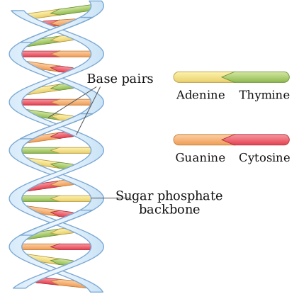
The Watson–Crick DNA double helix has two antiparallel strands coiled around a common axis, with complementary base pairs stacked inside.
As this platform is text-based, here is a fully labelled description of the diagram (the structural features a hand-drawn double helix must show):
- Two polynucleotide chains, coiled around a common central axis in a right-handed double helix.
- The two strands are antiparallel — one runs 5'→3' while the other runs 3'→5'.
- The sugar (deoxyribose)–phosphate backbone runs along the outside of the helix on both strands, giving the molecule its structural framework.
- The nitrogenous bases (adenine, guanine, cytosine, thymine) project inward, perpendicular to the axis, and pair with a base on the opposite strand via hydrogen bonds — this is complementary base pairing: Adenine pairs with Thymine via 2 hydrogen bonds (A=T), and Guanine pairs with Cytosine via 3 hydrogen bonds (G≡C).
- The two strands, held together by base-pair hydrogen bonds and base stacking, form a helix with a diameter of about 20 Å (2 nm).
- Adjacent base pairs are separated by about 3.4 Å, and a complete turn of the helix spans about 34 Å, giving roughly 10 base pairs per turn. …
Unlock everything free for 14 days
- Full step-by-step solutions
- Concept-first explanations
- Methods, shortcuts & mistakes
- PYQ mapping + timed mock tests
Full access for 14 days. No credit card required.