Q.Draw the different types of human WBC.
You're viewing a preview — the full solution, concept, methods & PYQ mapping are locked.
Start your 14-day free trial to unlock the full solution →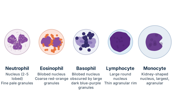
Human WBCs (leukocytes) are of five types, divided into granulocytes (Neutrophils, Eosinophils, Basophils) and agranulocytes (Lymphocytes, Monocytes); a hand-drawn figure isn't possible in this format, so each type's defining shape and staining features are described here for you to sketch and label yourself.
White blood cells are classified based on whether their cytoplasm shows visible granules under the microscope after staining:
Granulocytes (granular cytoplasm, lobed nucleus):
- Neutrophils: the most abundant WBC; nucleus has 2-5 lobes connected by thin strands (polymorphonuclear); cytoplasm shows fine, pale (neutral-staining) granules. Function: phagocytose bacteria, first responders in infection.
- Eosinophils: nucleus typically bilobed; cytoplasm packed with large, coarse granules that stain reddish-orange with eosin. Function: combat parasitic infections and involved in allergic responses.
- Basophils: least abundant WBC; nucleus is bilobed but often obscured by very large, coarse granules that stain dark blue-purple with basic dyes. Function: release histamine and heparin, involved in inflammatory/allergic responses.
Agranulocytes (no visible granules, rounder nucleus):
4. Lymphocytes: small to medium cells with a large, round, densely-stained nucleus that fills most of the cell, leaving only a thin rim of clear cytoplasm. Function: B-lymphocytes (antibody production) and T-lymphocytes (cell-mediated immunity). …
Unlock everything free for 14 days
- Full step-by-step solutions
- Concept-first explanations
- Methods, shortcuts & mistakes
- PYQ mapping + timed mock tests
Full access for 14 days. No credit card required.