Q.Draw and label the parts of a typical Chordate.
You're viewing a preview — the full solution, concept, methods & PYQ mapping are locked.
Start your 14-day free trial to unlock the full solution →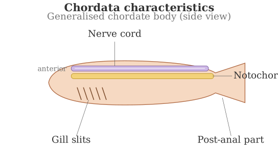
A typical chordate body plan is defined by four features present at some stage of the life cycle — notochord, dorsal hollow nerve cord, paired pharyngeal gill slits, and a post-anal tail — arranged along the body's antero-posterior axis; we describe and label these here in words, since a hand-drawn diagram cannot be produced in this format.
Picture a simple elongated chordate body drawn horizontally, with the head (anterior) end on the left and the tail (posterior) end on the right. Along its length, four structures should be labelled:
-
Notochord — a firm, flexible, rod-like structure running the length of the body just beneath the dorsal nerve cord, providing axial support; it is present at least during the embryonic stage in all chordates (replaced by the vertebral column in most adult vertebrates).
-
Dorsal, tubular (hollow) nerve cord — lies above (dorsal to) the notochord, running along the back; it is single and hollow (fluid-filled), unlike the solid, ventral, paired nerve cords of most invertebrates, and develops into the brain and spinal cord in vertebrates.
-
Paired pharyngeal gill slits — a series of paired openings in the wall of the pharynx (throat region), connecting the pharynx to the exterior; they function in filter-feeding and/or respiration in aquatic chordates, and though they may only appear in the embryo of terrestrial vertebrates, they are present at some life stage in every chordate.
…
Unlock everything free for 14 days
- Full step-by-step solutions
- Concept-first explanations
- Methods, shortcuts & mistakes
- PYQ mapping + timed mock tests
Full access for 14 days. No credit card required.