Question 71 of 99
Q.Draw the structure of T.S. of mature anther and label its parts.
Tamil Nadu DgeTamil Nadu HSC (DGE) Board 2020Subjective· 3mImportance★★★★★
72% · 71/99 Questions
You're viewing a preview — the full solution, concept, methods & PYQ mapping are locked.
Start your 14-day free trial to unlock the full solution →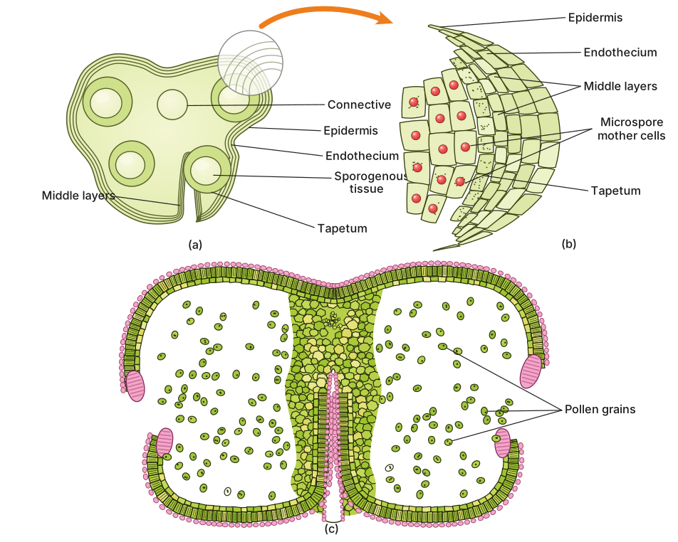
A T.S. of a mature anther shows two lobes joined by a connective, each lobe with two pollen sacs bounded by a four-layered wall (epidermis, endothecium, middle layers, tapetum) enclosing pollen grains.
Since a diagram cannot be rendered here, the structure of a T.S. of a mature anther is described part by part, as it would be labelled:
- Overall shape: in transverse section, the anther is typically tetragonal (four-sided/four-lobed in outline) and bilobed, i.e. it consists of two theca (lobes) held together in the middle.
- Connective: the central sterile tissue that joins the two lobes of the anther; it contains a vascular strand (vascular bundle) that supplies nutrients to the developing pollen sacs.
- Microsporangia (pollen sacs): each lobe (theca) of the anther contains two microsporangia, so a mature anther typically has four microsporangia (pollen sacs) in total, arranged in two pairs, one pair per lobe.
- Anther wall (from outside to inside): each microsporangium is bounded by a wall of four distinct layers --
- Epidermis: the outermost, single-layered protective covering.
- Endothecium: the layer just beneath the epidermis; its cells develop fibrous (radial) thickenings that help the anther dehisce (split open) at maturity to release pollen.
- Middle layers: 2-3 thin-walled layers lying between the endothecium and the tapetum; they get crushed and disappear as the anther matures. …
Unlock everything free for 14 days
- Full step-by-step solutions
- Concept-first explanations
- Methods, shortcuts & mistakes
- PYQ mapping + timed mock tests
Full access for 14 days. No credit card required.