Q.Describe the internal structure of a Dicot root.
You're viewing a preview — the full solution, concept, methods & PYQ mapping are locked.
Start your 14-day free trial to unlock the full solution →A dicot root (TS) shows epiblema → cortex → endodermis (Casparian strips, passage cells) → pericycle → radial vascular bundles with exarch xylem (usually 2–4, tetrarch) alternating with phloem, conjunctive tissue between them, and little/no pith.
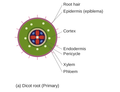
The internal (transverse-section) structure of a young dicot root is described from the outside inward — an 8-mark topic in the NCERT/CBSE-aligned TS Intermediate anatomy-of-flowering-plants unit.
Diagram (described): draw a circular transverse section. From the outermost ring inward, label: epiblema (with a few root hairs) → broad cortex → single-layered endodermis → pericycle → the central vascular region showing star-shaped xylem arms reaching outward (exarch) with phloem patches lying in between, conjunctive tissue separating xylem and phloem, and a small or absent pith at the centre.
1. Epiblema (piliferous layer / epidermis):
- The outermost single layer. It has no cuticle and no stomata, and many of its cells extend outward as unicellular root hairs that absorb water and minerals. It is protective and absorptive.
2. Cortex:
- A wide zone of many layers of thin-walled parenchyma cells with intercellular spaces. It stores food and conducts absorbed water inward towards the vascular tissue. (There is no distinct hypodermis in the root.)
3. Endodermis:
- The innermost single layer of the cortex, made of barrel-shaped cells fitting closely without spaces. Their radial and inner walls bear band-like Casparian strips (thickenings of suberin/lignin) that regulate water movement.
- Opposite the protoxylem (xylem arms), some thin-walled cells called passage cells (transfusion cells) remain unthickened to allow water to pass into the xylem.
4. Pericycle:
- A single (sometimes few) layer(s) of parenchymatous cells just inside the endodermis. It is important because lateral (secondary) roots arise from the pericycle, and it also takes part in the formation of the vascular cambium during secondary growth.
5. Vascular bundles (stele):
- Arranged in a radial fashion — i.e. xylem and phloem lie on separate radii, alternating with each other (not on the same radius as in stems).
- The xylem is exarch — the protoxylem points outward (towards the periphery) and metaxylem lies towards the centre. …
Unlock everything free for 14 days
- Full step-by-step solutions
- Concept-first explanations
- Methods, shortcuts & mistakes
- PYQ mapping + timed mock tests
Full access for 14 days. No credit card required.