Chemistry · Ch 2 — Structure of Atom
Wave Nature of Electromagnetic Radiation
Wave Nature of Electromagnetic Radiation
The Discovery of Electromagnetic Waves
In the mid-1800s, physicists were deeply puzzled by the nature of thermal radiation — the heat and light emitted by hot objects. They knew something was being radiated, but what was it? The answer came from James Clerk Maxwell in the 1870s. Maxwell developed a unified theory of electricity and magnetism, showing that an accelerating electric charge creates disturbances in the surrounding electric and magnetic fields. These disturbances travel outward as waves — electromagnetic waves.
Maxwell’s theory was a breakthrough. It predicted that light itself is an electromagnetic wave, a radical idea at the time. Heinrich Hertz later confirmed this experimentally. So, what we call electromagnetic radiation is simply the propagation of energy through space in the form of oscillating electric and magnetic fields.
The Nature of Electromagnetic Waves
An electromagnetic wave is not a physical wave like a sound wave or a water wave. It is a self-sustaining oscillation of two fields — an electric field and a magnetic field — that regenerate each other as the wave travels.
Unlike mechanical waves, electromagnetic waves do not need a medium. They can travel perfectly well through a vacuum. This is why we can see light from the Sun across the empty space between us.
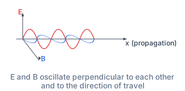
The figure in the textbook (Fig. 2.6) shows a simplified picture.
Drawn by us to help you understand the concept clearly, and verified to make sure it's accurate. For exams, practice from your textbook's own diagram.
Fig. 2.6 shows a single electromagnetic wave travelling along the x-axis (labelled with the direction of propagation). Two sinusoidal curves are drawn: the red curve is the electric field component E, oscillating along the vertical E-axis; the blue curve is the magnetic field component B, drawn oscillating along the tilted B-axis to suggest the third dimension — the standard flat projection of a 3-D wave. The two curves have the same wavelength (the distance between successive crests along x), the same frequency, the same amplitude (the maximum displacement from the x‑axis), and they are in phase — meaning that at any given point along x, the electric and magnetic fields reach their maxima and minima together.
The key physical idea is that an electromagnetic wave consists of two mutually perpendicular fields that are both perpendicular to the direction the wave travels. The electric field vibrates in one plane, the magnetic field in a plane at right angles to it, and the wave itself moves along the line perpendicular to both. This is why the figure places the red and blue sinusoids in orthogonal planes, with the x‑axis as the common direction of travel.
In an electromagnetic wave, , , and the direction of propagation are all mutually perpendicular. This is a transverse wave — the oscillations are perpendicular to the direction of energy transfer.
The textbook uses this picture to introduce the fundamental wave relation. For any electromagnetic radiation in vacuum, the speed , frequency , and wavelength are linked by:
Here (the speed of light in vacuum), is the number of wave crests passing a fixed point per second (unit: hertz, Hz, or s), and is the distance between successive crests (unit: metre). The figure makes it clear that is measured along the x‑axis, between two consecutive points where the wave repeats itself.
A second quantity, the wavenumber , is also defined from this figure’s geometry:
Its SI unit is m, but in spectroscopy it is often given in cm. The wavenumber tells you how many wavelengths fit into a unit length — a larger wavenumber means a shorter wavelength.
Do not confuse the wavenumber (with a bar) with frequency . They are different quantities with different units. The figure’s wavelength is the link between them: and . …
The electric field () and the magnetic field () are perpendicular to each other, and both are perpendicular to the direction in which the wave is moving. This makes electromagnetic waves transverse waves.
Four Key Properties of Electromagnetic Radiation
The textbook lists four essential properties. Each one is important for understanding how light and other radiations behave.
Property (i): Mutual Perpendicularity
The oscillating electric field and the oscillating magnetic field are perpendicular to each other. Furthermore, both fields are perpendicular to the direction of propagation of the wave. In the simplified diagram, if the wave moves to the right, the electric field might oscillate up and down (vertical plane), while the magnetic field oscillates in and out of the page (horizontal plane). All three directions — electric field, magnetic field, and wave travel — are mutually perpendicular.
Property (ii): No Medium Required
Electromagnetic waves can travel through a vacuum. Sound waves need air (or some other material) to compress and rarefy; water waves need water. But electromagnetic waves are disturbances in the fields themselves, and fields exist everywhere in space, even in a vacuum. This is a fundamental difference.
Property (iii): The Electromagnetic Spectrum
There are many types of electromagnetic radiation, and they differ from one another in their wavelength (or frequency). The entire collection of these radiations, arranged by wavelength or frequency, is called the electromagnetic spectrum.
The spectrum is divided into regions, each with a name and typical uses:
| Region | Approximate Frequency (Hz) | Common Use / Source |
|---|---|---|
| Radio waves | Broadcasting (radio, TV) | |
| Microwaves | Radar, microwave ovens | |
| Infrared (IR) | Heating, thermal imaging | |
| Visible light | What our eyes can detect | |
| Ultraviolet (UV) | Component of sunlight, causes tanning | |
| X-rays | Medical imaging | |
| Gamma rays | Radioactive decay, cancer treatment |
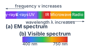
The visible region is only a tiny sliver of the entire spectrum, roughly from Hz to Hz. Our eyes are sensitive only to this narrow band. Special instruments (like radio telescopes, infrared cameras, or X-ray detectors) are needed to see the rest.
Drawn by us to help you understand the concept clearly, and verified to make sure it's accurate. For exams, practice from your textbook's own diagram.
What Fig. 2.7 Actually Shows
The figure is split into two parts. Part (a) is a horizontal bar representing the entire electromagnetic spectrum, divided into coloured segments from left to right: gamma rays, X-rays, ultraviolet (UV), a narrow visible band, infrared (IR), microwaves, and radio waves. Two arrows orient the bar: frequency increases to the left, wavelength increases to the right. (The book's own version additionally prints logarithmic /Hz and /m scales spanning about – Hz and – m.) The visible region is a tiny sliver in the middle.
Part (b) is an expanded view of that visible sliver — a colour bar stretching from violet at 400 nm on the left to red at 750 nm on the right. The wavelength scale is marked in nanometres (nm). This is the only part of the entire spectrum that the human eye can detect.
The axes are logarithmic. Each step to the left multiplies the frequency by a factor of 10 (and divides the wavelength by 10). That is why gamma rays ( m) and radio waves ( m) can sit on the same diagram.
The Physical Idea
The figure drives home one central point: visible light is not special — it is just a tiny slice of a much larger continuum of electromagnetic radiation. All these waves, from gamma rays to long radio waves, are the same kind of physical entity: oscillating electric and magnetic fields that travel at the same speed in vacuum. They differ only in their frequency and wavelength .
The textbook uses this figure to introduce the inverse relationship between frequency and wavelength. As you move left to right across the spectrum, frequency drops by 24 orders of magnitude while wavelength rises by 24 orders of magnitude. The visible region sits near the middle, with frequencies around Hz and wavelengths around m.
The Key Formula
The single equation that ties the whole figure together is the wave relation:
where:
- is the speed of light in vacuum, m s (precisely m s)
- (Greek letter nu) is the frequency in hertz (Hz, or s) — the number of wave crests passing a point per second
- (Greek letter lambda) is the wavelength in metres (m) — the distance between successive crests
Because is constant, if you know either or , you can calculate the other. For example, the textbook works out that violet light at 400 nm has a frequency of Hz, while red light at 750 nm has a frequency of Hz. The product in both cases equals m s.
Do not confuse the symbol (nu) with the letter v. In handwriting, many students write looking like a v, and then mistakenly treat it as velocity. The velocity here is , not .
A second quantity introduced alongside this figure is the wavenumber (nu bar), defined as the number of wavelengths per unit length:
Its SI unit is m, but in practice spectroscopists often use cm. The wavenumber is proportional to frequency (since ), so it is a convenient way to express the same information on a linear scale.
What the Figure Teaches You to Do
When you look at Fig. 2.7, you should be able to: …
Property (iv): Characterising Radiation — Frequency, Wavelength, and Wavenumber
Electromagnetic radiation is described by two fundamental quantities:
- Frequency (): The number of complete waves that pass a fixed point in one second. The SI unit is hertz (Hz), which is the same as (per second). It is named after Heinrich Hertz.
- Wavelength (): The distance between two successive crests (or troughs) of a wave. The SI unit is the metre (m). Because many electromagnetic waves have very small wavelengths, smaller units like nanometres (nm, ) or ångströms (Å, ) are often used.
A third quantity, the wavenumber (), is also commonly used in spectroscopy. It is defined as the number of wavelengths per unit length.
The SI unit of wavenumber is , but the unit (reciprocal centimetre) is very common in practice.