Biology · Ch 2 — Human Reproduction
Fertilisation and Implantation
Fertilisation and Implantation
The Journey of the Sperm and the Egg
Fertilisation is the fusion of a sperm with an ovum to form a single-celled zygote. In humans, this event takes place in the ampullary region of the fallopian tube, close to the isthmus. The ovum released from the ovary is carried into the fallopian tube by the fimbriae, and it is here that it waits for a sperm.
The process is not instantaneous. Millions of sperm are deposited in the vagina during copulation, but only a few hundred reach the site of fertilisation. The journey is a brutal selection process. Sperm must swim through the cervix, cross the uterus, and enter the fallopian tube. Along the way, secretions from the female reproductive tract capacitate the sperm — a series of physiological changes that make the sperm capable of penetrating the egg. Without capacitation, fertilisation cannot occur.
The Moment of Fusion
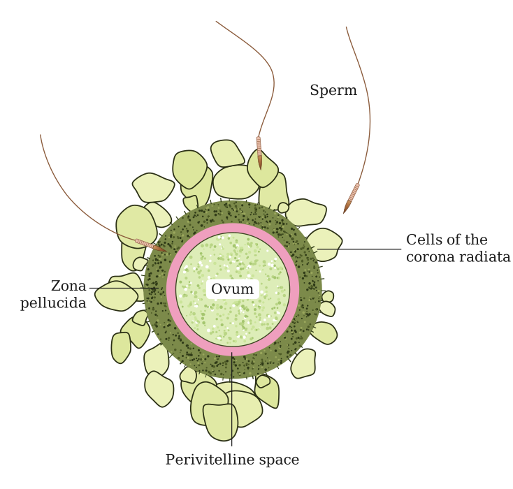
When a capacitated sperm reaches the ovum, it must first penetrate the two protective layers surrounding the egg: the outer corona radiata and the inner zona pellucida. The sperm releases enzymes from its acrosome (the cap-like structure at its head) to digest these layers. This is called the acrosomal reaction.
Once a single sperm makes contact with the egg's plasma membrane, the egg immediately releases cortical granules that harden the zona pellucida. This is the zona reaction, and it prevents any other sperm from entering — a critical block to polyspermy. The nuclei of the sperm and egg then fuse, and the haploid sets of chromosomes combine to restore the diploid number (46 chromosomes in humans). The fertilised egg is now called a zygote.
The First Cell Division and the Journey to the Uterus
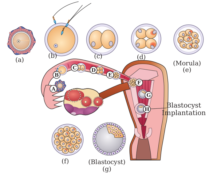
The zygote is a single cell, but it does not stay that way for long. It begins to divide by mitosis as it is slowly transported toward the uterus by the peristaltic movements of the fallopian tube and the beating of its cilia. The first division produces two cells, then four, then eight, and so on. These early cells are called blastomeres.
By the time the embryo reaches the uterus, it has become a solid ball of 16–32 cells called a morula. The morula continues to divide and soon develops a fluid-filled cavity. At this stage, it is called a blastocyst.
The Blastocyst: Structure and Specialisation
The blastocyst is a highly organised structure. It has two distinct cell populations:
- Trophoblast: The outer layer of cells. These cells will later form the placenta and other embryonic membranes. They do not contribute to the body of the embryo itself.
- Inner cell mass: A cluster of cells attached to the trophoblast on one side. This mass will develop into the actual embryo.
The blastocyst remains free-floating in the uterine cavity for a few days. During this time, the zona pellucida (the protective coat that surrounded the egg) degenerates, exposing the trophoblast directly to the uterine lining.
Implantation: Anchoring the Embryo
Implantation is the process by which the blastocyst attaches to and embeds itself into the endometrium — the inner lining of the uterus. This occurs approximately 7 days after fertilisation.
The trophoblast cells are the key players here. They secrete enzymes that digest the endometrial tissue, allowing the blastocyst to burrow into the thick, blood-rich lining. The endometrium, under the influence of progesterone from the corpus luteum, has become highly vascularised and secretory, ready to nourish the embryo.
Once implanted, the trophoblast cells continue to proliferate and differentiate. They send out finger-like projections called chorionic villi that grow into the endometrium. These villi, along with the underlying uterine tissue, form the placenta — the organ that will sustain the embryo for the rest of pregnancy.
The placenta is a temporary organ that is derived from both fetal tissue (chorionic villi) and maternal tissue (endometrium). It is the interface for all exchange between mother and fetus.
The Timeline of Early Development
| Stage | Location | Key Event |
|---|---|---|
| Fertilisation | Ampullary region of fallopian tube | Sperm fuses with ovum; zygote formed |
| Cleavage (cell division) | Fallopian tube | Zygote divides into blastomeres; morula forms |
| Blastocyst formation | Uterine cavity | Fluid-filled cavity appears; inner cell mass and trophoblast differentiate |
| Implantation | Endometrium of uterus | Blastocyst attaches and embeds; trophoblast invades endometrium |
What Happens After Implantation? …
Drawn by us to help you understand the concept clearly, and verified to make sure it's accurate. For exams, practice from your textbook's own diagram.
This figure gives you a close-up, almost microscopic view of the moment just before fertilisation. At the centre is the ovum (the secondary oocyte), a large, round cell. Wrapped tightly around it is a thick, translucent layer called the zona pellucida — you can think of it as the ovum's protective coat. Outside the zona pellucida, the figure shows a ring of corona radiata cells, which are follicle cells that still cling to the ovum after ovulation.
Scattered around the outside of the corona radiata are several sperms. The figure captures them at the stage where they have already travelled through the female reproductive tract and are now swarming around the ovum. Some sperms are shown with their heads pressed against the corona radiata cells, while others have already penetrated through that layer and are making direct contact with the zona pellucida. …
Drawn by us to help you understand the concept clearly, and verified to make sure it's accurate. For exams, practice from your textbook's own diagram.
The figure traces the journey of the female gamete and the early embryo through the female reproductive tract. It shows the ovary, the fallopian tube (divided into infundibulum, ampulla, and isthmus), and the uterus in a single continuous diagram. The ovary is on the left, releasing an ovum into the peritoneal cavity near the fimbriae of the infundibulum.
The key event shown is fertilisation, which occurs in the ampullary region of the fallopian tube — the widest, longest part of the tube. From that point, the figure follows the developing embryo as it moves through the isthmus toward the uterus. Along this path, the diagram labels the successive cleavage stages: zygote (the single-celled fertilised egg), then 2-cell, 4-cell, and 8-cell stages, followed by morula (a solid ball of about 16–32 cells), and finally blastocyst (a hollow, fluid-filled structure with an inner cell mass). Each stage is placed at a progressively more uterine position along the tube.
Arrows in the figure indicate the direction of movement — from the ovary, through the tube, and into the uterine cavity. The blastocyst is shown approaching the uterine wall, where implantation will occur. The figure does not depict implantation itself, only the embryo's arrival at the uterus.
The figure makes clear that fertilisation happens in the ampulla, not in the uterus or ovary. The entire journey from fertilisation to the blastocyst stage takes about 5–7 days, during which the embryo is moving through the fallopian tube. …