Biology · Ch 2 — Human Reproduction
Pregnancy and Embryonic Development
Pregnancy and Embryonic Development
The journey from fertilisation to the birth of a fully formed baby is called pregnancy, and the process by which the baby grows inside the mother is embryonic development. This section traces that entire timeline, from the moment a zygote is formed to the moment a child is delivered.
From Zygote to Blastocyst
After fertilisation in the ampulla of the fallopian tube, the single-celled zygote immediately begins to divide by mitosis — a process called cleavage. These divisions are rapid, and the cells (now called blastomeres) get smaller with each division without increasing the overall size of the embryo. The embryo moves down the fallopian tube toward the uterus as it divides.
By the time it reaches the 8- to 16-cell stage, the embryo is called a morula (from the Latin for mulberry, which it resembles). The morula continues to divide and, as it enters the uterine cavity, it transforms into a blastocyst. A blastocyst has two distinct parts:
- An outer layer of cells called the trophoblast.
- An inner group of cells called the inner cell mass.
The trophoblast will later form the placenta, while the inner cell mass will develop into the actual embryo.
Implantation
Once the blastocyst reaches the uterus, it needs to attach itself to the uterine lining to continue growing. This attachment is called implantation. The trophoblast cells secrete enzymes that digest the endometrial tissue, allowing the blastocyst to embed itself into the endometrium. Implantation is completed by about the 7th day after fertilisation.
After implantation, the uterine endometrium undergoes further changes and is now called the decidua. The trophoblast cells also begin to develop finger-like projections called chorionic villi, which grow into the endometrium. Together, the chorionic villi and the surrounding uterine tissue form the placenta.
The Placenta: A Lifeline
The placenta is a temporary organ that connects the developing embryo to the mother's uterine wall. It serves as the interface for the exchange of nutrients, oxygen, and waste products between the mother and the foetus. The placenta performs several critical functions:
- Supply: It delivers oxygen and nutrients (like glucose, amino acids, and vitamins) from the mother's blood to the foetus.
- Removal: It carries away carbon dioxide and nitrogenous wastes (like urea) from the foetal blood to the mother's blood for excretion.
- Barrier: It prevents many harmful substances and pathogens from reaching the foetus, though it is not a perfect barrier (some viruses and drugs can cross it).
- Hormone production: The placenta acts as an endocrine gland. It secretes several hormones essential for maintaining pregnancy, including:
- Human chorionic gonadotropin (hCG): This hormone is produced by the trophoblast cells soon after implantation. It maintains the corpus luteum in the ovary, which continues to secrete progesterone and estrogen to keep the uterine lining intact. hCG is the hormone detected in pregnancy tests.
- Human placental lactogen (hPL): Also called human chorionic somatomammotropin, this hormone helps regulate maternal metabolism and promotes the growth of the mammary glands.
- Estrogens and Progesterone: These are produced in large quantities by the placenta after the first trimester. They are crucial for maintaining the pregnancy, suppressing ovulation, and preparing the breasts for lactation.
The foetus is connected to the placenta by the umbilical cord, which contains two umbilical arteries (carrying deoxygenated blood and wastes from the foetus to the placenta) and one umbilical vein (carrying oxygenated blood and nutrients from the placenta to the foetus).
The Three Trimesters of Pregnancy
Human pregnancy lasts about nine months (approximately 40 weeks from the last menstrual period) and is divided into three trimesters of three months each.
The developing individual is called an embryo for the first eight weeks after fertilisation. After that, it is called a foetus until birth.
| Trimester | Duration | Key Developments |
|---|---|---|
| First | 0–3 months | Major organ systems begin to form (organogenesis). The heart starts beating. The embryo is most vulnerable to damage from drugs, alcohol, and infections. By the end, the foetus is about 5–8 cm long and can move its limbs. |
| Second | 3–6 months | The foetus grows rapidly. Limbs, fingers, and toes are fully formed. The mother can feel foetal movements ("quickening"). The foetus's skin is covered with a fine hair called lanugo and a waxy coating called vernix caseosa. By the end, the foetus is about 30 cm long. |
| Third | 6–9 months | The foetus continues to grow and mature. The lungs and brain develop significantly. The foetus gains weight and stores fat. It usually turns head-down in preparation for birth. By the end, the foetus is about 50 cm long and weighs around 3–3.5 kg. |
Parturition and Lactation …
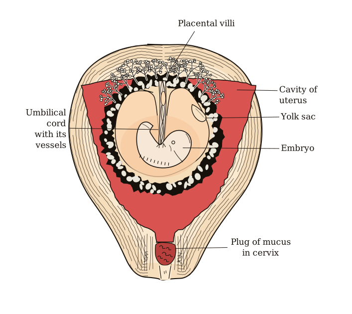
Drawn by us to help you understand the concept clearly, and verified to make sure it's accurate. For exams, practice from your textbook's own diagram.
The figure shows a cross-section of the human uterus during the later stages of pregnancy, with a fully developed foetus inside. The uterine wall is drawn as a thick, layered structure, and the foetus is positioned within the uterine cavity, curled in the typical foetal posture. The most prominent feature is the umbilical cord, a long, rope-like structure that connects the foetus’s navel to the placenta. The placenta itself is shown as a disc-shaped organ attached to the inner lining of the uterine wall.
At the interface between the placenta and the uterus, the figure highlights chorionic villi — finger-like projections of foetal tissue that extend into the uterine tissue. These villi are shown interdigitating (interlocking) with the maternal uterine tissue, creating a large surface area for exchange. The uterine wall is labelled to indicate the endometrium (the inner lining) and the myometrium (the thick muscular layer beneath it). The foetus is surrounded by the amniotic cavity, filled with amniotic fluid, which cushions and protects the foetus.
Arrows in the figure typically indicate the direction of nutrient and gas exchange across the placenta: oxygen and nutrients move from maternal blood in the uterine tissue into the foetal blood in the chorionic villi, while carbon dioxide and waste products move in the opposite direction. The figure does not show any additional panels, dates, or artefacts — it is a single, labelled diagram of the foetus in utero. …