Biology · Ch 2 — Human Reproduction
Gametogenesis
Gametogenesis
The Purpose of Gametogenesis
Reproduction in humans requires the formation of specialised sex cells — the male gamete (sperm) and the female gamete (ovum). The process by which these haploid gametes are produced from the diploid germ cells in the gonads is called gametogenesis. It is the foundation of sexual reproduction, ensuring that when a sperm and an ovum fuse during fertilisation, the resulting zygote has the correct diploid number of chromosomes.
Two Distinct Processes
Gametogenesis is not a single process; it occurs differently in males and females. In males, it is called spermatogenesis and takes place in the testes. In females, it is called oogenesis and takes place in the ovaries. Both processes involve meiosis (reduction division) to halve the chromosome number, but they differ significantly in timing, duration, and the number of functional gametes produced.
Spermatogenesis: The Formation of Sperm
Spermatogenesis begins at puberty and continues throughout a male's life. The entire process occurs within the seminiferous tubules of the testes.
The process starts with the spermatogonia (singular: spermatogonium), which are the immature male germ cells. These cells are diploid and are present on the inner wall of the seminiferous tubules. Spermatogonia undergo repeated mitotic divisions, increasing their numbers. Some of these spermatogonia stop dividing and begin to grow, transforming into primary spermatocytes.
Each primary spermatocyte is diploid and now enters the first meiotic division (meiosis I). This is a reductional division, meaning it halves the chromosome number. The result is two haploid cells called secondary spermatocytes.
The secondary spermatocytes then immediately undergo the second meiotic division (meiosis II), which is an equational division. Each secondary spermatocyte divides to form two haploid spermatids. So, from one primary spermatocyte, we get four spermatids.
The spermatids are small, haploid cells, but they are not yet functional sperm. They undergo a dramatic transformation called spermiogenesis (or spermateliosis). During this process, the spermatid changes shape: it develops a head, a middle piece, and a tail. The nucleus condenses, the Golgi apparatus forms the acrosome (a cap-like structure containing enzymes), and mitochondria gather in the middle piece to provide energy for movement.
Finally, the mature spermatozoa (sperm) are released from the Sertoli cells (also called nurse cells) into the lumen of the seminiferous tubules. The Sertoli cells provide nourishment and support to the developing germ cells throughout spermatogenesis.
The entire duration of spermatogenesis in humans takes approximately 64 days.
Oogenesis: The Formation of the Ovum
Oogenesis is a markedly different process. It begins during the embryonic development of a female and is completed only after fertilisation. It occurs in the ovaries.
The process starts with the oogonia (singular: oogonium), diploid female germ cells that form and multiply by mitosis during the embryonic development of the female — no new oogonia are formed or added after birth. These cells then enter the first meiotic division (prophase I) and get temporarily arrested at that stage; it is this arrested state that defines a cell as a primary oocyte. Each primary oocyte is diploid and is surrounded by a layer of granulosa cells, and together they form a structure called a primary follicle. The arrest lasts until the female reaches puberty — a key difference from spermatogenesis, which only starts at puberty.
At puberty, a small number of primary follicles (about 60,000–80,000) are present in each ovary. With each menstrual cycle, a few of these follicles begin to grow. Typically, only one follicle fully matures. The primary oocyte inside this mature follicle resumes its first meiotic division. This division is unequal: it produces one large haploid cell called the secondary oocyte and a very small, non-functional cell called the first polar body. The first polar body may or may not undergo a second meiotic division.
The secondary oocyte now begins the second meiotic division but gets arrested again — this time at metaphase II. It is released from the ovary during ovulation in this arrested state.
The second meiotic division is only completed if the secondary oocyte is fertilised by a sperm. Upon fertilisation, the secondary oocyte completes meiosis II, again unequally. This produces a large, mature ovum (or egg) and a second polar body. The first polar body may also divide to form two polar bodies. In total, one primary oocyte yields one functional ovum and up to three polar bodies, which eventually degenerate.
A common mistake is to think that the secondary oocyte is the mature ovum. It is not. The secondary oocyte is released at ovulation and only becomes the mature ovum after fertilisation completes meiosis II.
A Summary Comparison
| Feature | Spermatogenesis | Oogenesis |
|---|---|---|
| Location | Seminiferous tubules of testes | Ovaries |
Drawn by us to help you understand the concept clearly, and verified to make sure it's accurate. For exams, practice from your textbook's own diagram.
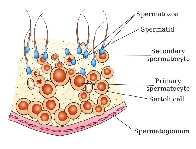
This figure is a magnified, wedge-shaped slice through the wall of a seminiferous tubule. The tubule is a long, coiled tube inside the testis, and its wall is the site where sperm are made. The diagram is oriented so that the outer edge of the wedge (the basement membrane side) is at the top or left, and the inner edge (the lumen, or central hollow space) is at the bottom or right.
The key structural feature is the basement membrane, a thin, non-cellular layer that forms the outer boundary of the tubule wall. Just inside this membrane lies a layer of spermatogonia — the diploid stem cells that divide by mitosis to maintain their own population and to produce cells that will undergo meiosis. These are the most immature germ cells, located at the periphery.
Moving inward toward the lumen, the figure shows the successive stages of spermatogenesis in a clear sequence. The spermatogonia give rise to primary spermatocytes (diploid, in prophase I of meiosis), which are larger and lie closer to the lumen. These then divide to form secondary spermatocytes (haploid, smaller, and short-lived), which are shown even closer to the lumen. The secondary spermatocytes quickly complete meiosis II to produce spermatids (haploid, round, and non-motile), which are positioned near the lumen's edge. Finally, the spermatids undergo spermiogenesis (no further division, just differentiation) to become spermatozoa (mature sperm), which are shown lining the lumen, with their heads embedded in Sertoli cells and their tails projecting into the lumen.
Interspersed among all these germ cells are the Sertoli cells (also called sustentacular cells). These are tall, columnar cells that extend from the basement membrane all the way to the lumen. The figure shows them as large, irregularly shaped cells with a prominent nucleus. Their role is supportive: they provide nourishment to the developing germ cells, secrete fluid, and help in the release of mature sperm into the lumen. The diagram often uses arrows or a linear arrangement to indicate the direction of development — from the basement membrane outward toward the lumen — making it clear that spermatogenesis proceeds from the periphery to the center.
The figure does not show the interstitial cells (Leydig cells) because those lie outside the seminiferous tubule, in the connective tissue between tubules. The focus here is strictly on the cellular events within the tubule wall. …
Drawn by us to help you understand the concept clearly, and verified to make sure it's accurate. For exams, practice from your textbook's own diagram.
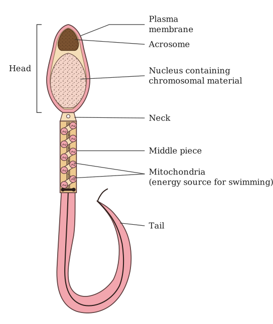
Figure 2.6 shows a single human sperm cell, drawn in a side view so that its entire length is visible. The cell is enclosed by a continuous plasma membrane, and the diagram is divided into four distinct regions from left to right: the head, the neck, the middle piece, and the tail.
The head is the most prominent part. It contains a compact, elongated haploid nucleus, which carries the paternal genetic material. Capping the front half of this nucleus is the acrosome — a specialised vesicle filled with hydrolytic enzymes that will later help the sperm penetrate the egg's outer layers. The head is not round; it is drawn as an oval that tapers slightly toward the front, reflecting the textbook's description of an "elongated" shape.
Immediately behind the head is a short, narrow neck. This region connects the head to the rest of the cell and contains the proximal centriole, which is important for the first mitotic division after fertilisation.
The middle piece follows the neck. It is thicker than the neck and is packed with mitochondria arranged in a spiral around the core of the flagellum. These mitochondria generate the ATP needed to power the tail's movement. The middle piece is the metabolic engine of the sperm.
Finally, the tail — also called the flagellum — extends from the middle piece to the far end. It is a long, slender structure that tapers gradually. The tail's rhythmic, whip-like beating propels the sperm forward through the female reproductive tract. …
Drawn by us to help you understand the concept clearly, and verified to make sure it's accurate. For exams, practice from your textbook's own diagram.
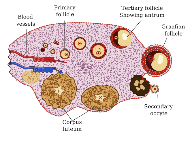
This figure is a cross-section of the ovary, cut to reveal its internal structure. The outer boundary is the germinal epithelium, and the bulk of the interior is the stroma — the connective tissue framework of the ovary. Embedded within the stroma, the figure shows a sequence of follicles at different stages of development, arranged roughly from the outer edge toward the centre.
The earliest stage shown is the primary follicle, located just beneath the surface. It consists of a primary oocyte surrounded by a single layer of flattened follicular cells. As development proceeds, the follicle enlarges and the follicular cells multiply into several layers, forming the secondary follicle. A fluid-filled cavity, the antrum, begins to appear within the follicular cells, marking the transition to the tertiary follicle. The antrum grows larger, pushing the oocyte to one side, and the follicle becomes the Graafian follicle — the mature, fully developed follicle ready for ovulation.
The figure then depicts ovulation: the Graafian follicle has ruptured, and a released ovum (the secondary oocyte) is shown being expelled from the ovary's surface. After ovulation, the ruptured follicle collapses and transforms into the corpus luteum, a yellowish glandular structure also labelled in the figure. The corpus luteum occupies a prominent position in the stroma, indicating its role in the post-ovulatory phase. …
Drawn by us to help you understand the concept clearly, and verified to make sure it's accurate. For exams, practice from your textbook's own diagram.
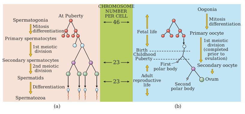
Figure 2.8 places spermatogenesis and oogenesis side by side as two vertical flowcharts, making the parallel and the key differences immediately visible. Each panel reads from top to bottom, following the cell as it moves through successive stages of development.
Panel (a) — Spermatogenesis starts at the top with a single spermatogonium (the diploid stem cell). An arrow leads downward to a primary spermatocyte, which is still diploid. The next arrow marks the first meiotic division, producing two secondary spermatocytes (now haploid). A second arrow shows the second meiotic division, which turns each secondary spermatocyte into two spermatids — so four haploid spermatids in total. The final arrow represents spermiogenesis (the transformation of spermatids into mature sperm), ending with four equal-sized sperms. The entire process is drawn as a single, unbranching chain: one cell becomes four identical gametes.
Panel (b) — Oogenesis begins with an oogonium (also diploid). The first arrow leads to a primary oocyte. Here the first meiotic division is shown as an unequal split: it produces one large secondary oocyte and one tiny first polar body. The second meiotic division is again unequal: the secondary oocyte divides into a large ovum and a small second polar body. Meanwhile, the first polar body may also divide (into two polar bodies), so the final result is one large ovum plus up to three small polar bodies. Unlike spermatogenesis, oogenesis yields only one functional gamete per starting cell, and the meiotic divisions are asymmetric — the cytoplasm is conserved in the egg. …