Botany · Ch 5 — Cell: The Unit of Life
Cilia and Flagella
Cilia and Flagella
Cilia and flagella are hair-like projections that extend outward from the surface of the cell, and both are involved in movement.
Cilia versus flagella
Both structures are outgrowths of the cell membrane, but they differ in size, number and the kind of movement they produce:
- Cilia (singular: cilium) are small, short structures. They work rather like oars, and their beating causes either the cell to move or the fluid surrounding the cell to move. Cilia are usually numerous and beat in a coordinated fashion.
- Flagella (singular: flagellum) are comparatively longer and fewer in number, and they are responsible for the movement of the cell itself.
Eukaryotic versus bacterial flagella
Prokaryotic bacteria also possess flagella, but a bacterial flagellum is structurally different from a eukaryotic flagellum. Although both are called by the same name and both drive movement, their internal construction is not the same, so they should not be treated as identical.
Internal structure — the axoneme and the 9+2 array
Electron microscopy shows that a cilium or flagellum is covered on the outside by the plasma membrane. Its central core is called the axoneme, and this contains a set of microtubules running parallel to the long axis of the structure. The arrangement of these microtubules is highly regular:
- There are nine doublets (pairs) of microtubules arranged radially around the outside — these are the peripheral microtubules.
- At the centre lies a pair of single microtubules — the central tubules.
Because of this pattern — nine outer doublets surrounding two central tubules — the arrangement is known as the 9+2 array.
Several connecting elements hold this array together:
- The two central tubules are joined to each other by bridges and are enclosed within a central sheath. …

Drawn by us to help you understand the concept clearly, and verified to make sure it's accurate. For exams, practice from your textbook's own diagram.
This is a stylised evocation of the electron micrograph of a cross-section through a cluster of cilia/flagella. Each roughly circular cross-section shows the same '9 + 2' arrangement of microtubules seen in the labelled diagram alongside it (panel b) -- nine peripheral doublets ringing a central pair -- as it actually appears un …
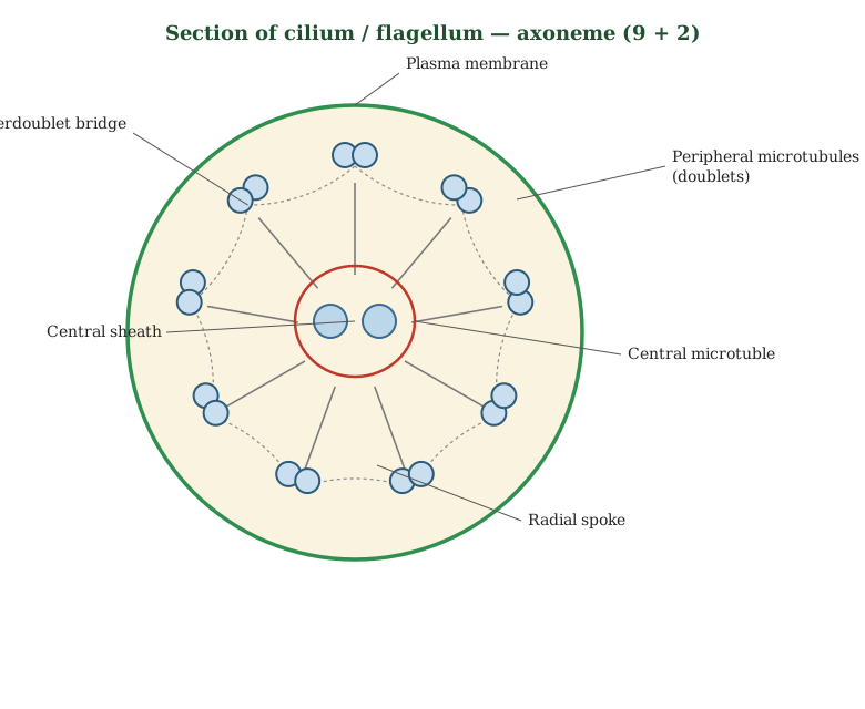
Drawn by us to help you understand the concept clearly, and verified to make sure it's accurate. For exams, practice from your textbook's own diagram.
This cross-section cuts across a cilium or flagellum to reveal the core skeleton, called the axoneme. Looking end-on, nine pairs of microtubules, known as doublets, are spaced evenly in a ring around the outside. At the very centre lies one extra pair of single microtubules. This nine-outer-doublets-plus-two-central pattern is described simply as the 9+2 arrangement and is found in nearly all cilia and flagella. Fine radial spokes reach inward from the doublets toward a central sheath that surrounds the middle pair, holding the whole bundle in order. The plasma membrane wraps the entire structure. The key idea is that this precis …