Q.Draw a neat diagram of enlarged view of one microsporangium of a Young anther of an angiosperm. Label the different layers of the wall and write the functions of the outermost and innermost layer.
You're viewing a preview — the full solution, concept, methods & PYQ mapping are locked.
Start your 14-day free trial to unlock the full solution →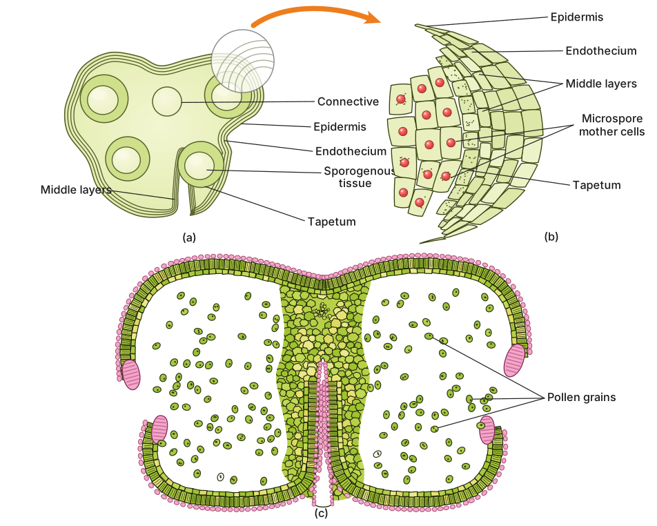
A young angiosperm microsporangium has four concentric wall layers — epidermis (protection), endothecium (dehiscence), middle layers (nutrition), and tapetum (nourishment of pollen) — surrounding the sporogenous tissue that will form pollen grains.
The microsporangium is the site where male gametophytes begin their journey in flowering plants. When you slice through a young anther and zoom in on one of its pollen sacs, you see a beautifully organized structure: concentric layers of cells wrapping around a central mass of cells destined to become pollen. This arrangement is not accidental — each layer has a specific role in nurturing and eventually releasing the pollen grains.
Structure of the Microsporangium Wall
From the outside moving inward, the microsporangium wall consists of four distinct layers:
1. Epidermis — The outermost protective jacket, a single layer of cells that shields the developing pollen from the external environment.
2. Endothecium — Just beneath the epidermis lies this layer of radially elongated cells. As the anther matures, the endothecium develops fibrous thickenings on its inner tangential and radial walls. These thickenings are hygroscopic (they respond to changes in moisture), and their differential expansion creates the mechanical force that splits the anther open at maturity, releasing the pollen. This process is called dehiscence.
3. Middle Layers — One to three layers of thin-walled cells sit between the endothecium and the tapetum. These layers are transient; they degenerate as the anther matures, contributing nutrients to the developing pollen grains.
4. Tapetum — The innermost layer, and arguably the most metabolically active. Tapetal cells are large, often multinucleate, and packed with dense cytoplasm. They nourish the developing microspore mother cells and later the pollen grains, secreting enzymes, hormones, and materials for the pollen wall (including sporopollenin, the remarkably resistant substance that forms the exine).
At the very center of this layered structure lies the sporogenous tissue, a group of compactly arranged homogeneous cells. Each of these cells has the potential to undergo meiosis and give rise to microspores, which will mature into pollen grains.
The tapetum is sometimes called the "nurse tissue" of the anther because it literally feeds the developing pollen. Without a functional tapetum, pollen development fails, leading to male sterility.
Functions of Key Layers
Epidermis (Outermost Layer):
Its primary function is protection. It forms a continuous barrier that prevents desiccation and shields the delicate internal tissues from mechanical injury and pathogen attack during the early stages of anther development.
Tapetum (Innermost Layer):
This layer is the nutritional powerhouse. Its functions include:
- Nourishing the developing microspore mother cells and microspores …
Unlock everything free for 14 days
- Full step-by-step solutions
- Concept-first explanations
- Methods, shortcuts & mistakes
- PYQ mapping + timed mock tests
Full access for 14 days. No credit card required.