Q.(a)
🔒You're viewing a preview — the full solution, concept, methods & PYQ mapping are locked.
🔒 Start your 14-day free trial to unlock the full solution →Part (a)Concept understanding — Male And Female Gametophyte
Let’s start with something you already know. Think of a plant like a mango tree. It produces flowers, and those flowers eventually turn into mangoes. How does that happen? The flower is the plant’s reproductive organ, and inside it, two tiny but crucial structures are made: the male gametophyte and the female gametophyte. These are not the “male” and “female” parts of the flower themselves (like the stamen and pistil) — they are the microscopic, single-generation cells that actually carry the genetic material to form the next plant.
The everyday intuition
Imagine you are baking a cake. The male gametophyte is like the packet of dry yeast — it’s small, mobile, and needs to be added to the mixture. The female gametophyte is like the bowl of flour, eggs, and sugar — it’s larger, stationary, and provides the environment where the yeast works. Without the yeast reaching the bowl, no cake rises. Without the bowl, the yeast has nothing to act on. In a flower, the male gametophyte (pollen grain) must travel to the female gametophyte (embryo sac) for fertilisation to happen.
The precise meaning
In NCERT biology, the male gametophyte is the pollen grain. It develops inside the anther (the top part of the stamen). A mature pollen grain contains two cells: a tube cell and a generative cell. The tube cell will grow a long tube down the style of the pistil, and the generative cell will divide to form two sperm cells. So the male gametophyte is not the whole stamen — it’s the tiny, single-celled (or two-celled) structure that carries the male genetic material.
The female gametophyte is the embryo sac. It develops inside the ovule, which is located in the ovary of the pistil. The embryo sac is a seven-celled, eight-nucleate structure (though you don’t need to memorise numbers for a prose subject — just know it’s a small, organised sac). It contains the egg cell (the female gamete) and other cells that help in fertilisation and nourishment. The female gametophyte is not the whole pistil — it’s the microscopic sac inside the ovule.
The male gametophyte (pollen grain) is haploid — it has only one set of chromosomes. The female gametophyte (embryo sac) is also haploid. When they fuse during fertilisation, they form a diploid zygote, which grows into the seed. This is why both are called “gametophytes” — they produce gametes (sperm and egg).
Why it matters …
Part (b)Concept understanding — Human Reproduction
Human Reproduction: A First Look
Think about what makes you you. You have your mother's eyes, your father's laugh, a mix of traits from two people you may never have met — your grandparents. That chain of life, stretching back thousands of years, runs through one process: human reproduction. It is not a topic for biology students alone. It is the story of how every single person on this planet came to be here.
The Big Picture: Why Does Reproduction Exist?
Every living thing has one fundamental drive: to continue its kind. A mango tree grows fruit so its seeds can sprout elsewhere. A bacterium splits in two. For humans, reproduction is more complex — and more personal. It is the reason families exist, why you have siblings or cousins, and why populations grow or shrink.
At its simplest, human reproduction is the biological process by which a new human individual is created. It requires two parents — a male and a female — each contributing a special cell that fuses to form a single, new cell. That single cell then grows, divides, and develops into a baby over about nine months.
Reproduction is not the same as sex. Sex is an act; reproduction is the entire sequence from cell fusion to birth. Not every act of sex leads to reproduction, and reproduction can sometimes happen with medical help (like IVF) without sex.
The Two Key Players: Male and Female Reproductive Systems
Your body, whether male or female, has a set of organs designed specifically for reproduction. These are not the same as the organs that keep you alive — they are the "spare set" dedicated to creating the next generation.
The Male System: Production and Delivery
The male reproductive system has one main job: to produce male sex cells (sperm) and deliver them to the female body. The key parts are:
- Testes (two oval organs in a pouch called the scrotum): These are the factories. They produce sperm continuously from puberty onwards — millions every day. They also produce testosterone, the hormone that drives male physical changes at puberty.
- Duct system (a series of tubes): Sperm travel from the testes through a long coiled tube (epididymis) where they mature, then through a muscular tube (vas deferens) that carries them toward the urethra.
- Accessory glands (seminal vesicles, prostate gland, bulbourethral glands): These add fluids to the sperm to nourish them and help them swim. The mixture of sperm and these fluids is called semen.
- Penis: The organ that delivers semen into the female reproductive tract.
The Female System: Production, Reception, and Nurturing
The female system has a bigger job: it produces female sex cells (eggs), receives sperm, and if fertilization occurs, houses and nourishes the developing baby for nine months. The key parts are:
- Ovaries (two almond-sized organs): These produce eggs — but unlike sperm, eggs are not made continuously. A female is born with all the eggs she will ever have (about 1–2 million at birth, reducing to around 300,000–400,000 by puberty). Each month, one egg matures and is released.
- Fallopian tubes (also called oviducts): These are not connected directly to the ovaries. When an egg is released, the finger-like ends of the tube sweep it inside. Fertilization — the meeting of sperm and egg — happens here, in the tube.
- Uterus (womb): A hollow, pear-shaped muscular organ. If fertilization happens, the embryo implants in the lining of the uterus and grows here. If not, the lining is shed — that is menstruation.
- Cervix: The lower, narrow part of the uterus that opens into the vagina. It produces mucus that changes consistency during the month to help or hinder sperm.
- Vagina: A muscular canal that receives the penis during intercourse and serves as the birth canal during delivery.
The female reproductive system has a monthly cycle (the menstrual cycle) that prepares the body for a possible pregnancy. This cycle is controlled by hormones and typically lasts about 28 days. The release of the egg (ovulation) happens around day 14. If no pregnancy occurs, the uterine lining is shed as menstrual blood — this is a normal, healthy process, not a "curse" or something to be ashamed of.
The Moment of Creation: Fertilization
When sperm from the male meet the egg from the female inside a fallopian tube, one sperm may penetrate the egg's outer layer. That single moment — fertilization — creates a new cell called a zygote. This zygote contains 23 chromosomes from the mother and 23 from the father, making 46 in total. That is the complete genetic blueprint for a new human being.
The zygote immediately begins to divide as it travels down the fallopian tube toward the uterus. By the time it reaches the uterus (about 5–7 days later), it has become a ball of about 100 cells called a blastocyst. This blastocyst burrows into the soft, thick lining of the uterus — a process called implantation. That is when pregnancy truly begins.
The Nine-Month Journey: Development
Once implanted, the developing human is called an embryo for the first eight weeks, and then a fetus from the ninth week until birth. During these months:
- A special organ called the placenta develops, connecting the mother's blood supply to the fetus. It delivers oxygen and nutrients and removes waste — without the mother's and baby's blood ever mixing directly.
- The umbilical cord links the baby to the placenta.
- The baby is protected by a fluid-filled sac (amniotic sac) that cushions it.
- All major organs form in the first three months (first trimester). The next three months (second trimester) are about growth and refinement. The final three months (third trimester) are about rapid weight gain and preparation for life outside the womb. …
Part (a)
- Role of stigma. The stigma is the receptive tip of the pistil. It recognises compatible pollen (through a chemical dialogue between pollen-wall proteins and stigma proteins), provides a sticky, sugary secretion for pollen to adhere and germinate, and rejects incompatible/self pollen — ensuring only suitable pollen forms a pollen tube.
- Post-pollination events → double fertilization. A 2-celled pollen grain (vegetative + generative cell) germinates; the vegetative cell forms the pollen tube through the style, and the generative cell divides to form two male gametes. Guided to the ovule, the tube enters via the micropyle, passes into a synergid and releases the two gametes. One gamete fuses with the egg (syngamy → diploid zygote); the other fuses with the two polar nuclei (triple fusion → triploid PEN/endosperm) — together this is double fertilization. …
Part (a): The stigma selectively accepts compatible pollen; the pollen tube then delivers two male gametes to the embryo sac, where one fuses with the egg and the other with the polar nuclei (double fertilization).
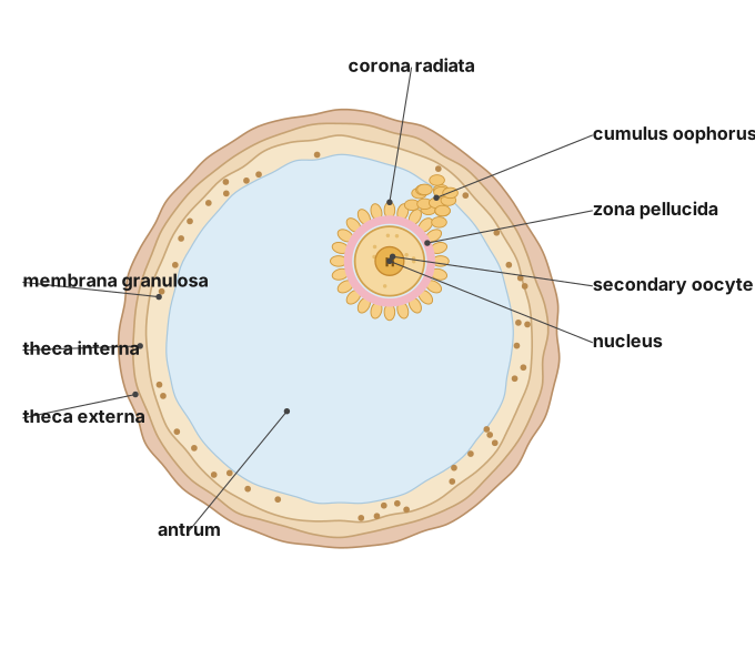
Part (b): The menstrual phase sheds the endometrium; the follicular phase is 'proliferative' because estrogen rebuilds the endometrium; at ovulation an LH surge ruptures the Graafian follicle to release the secondary oocyte, after which the corpus luteum forms.
Part (a)
Concept-first idea: Pollen–pistil interaction is a recognition process that lets a compatible pollen grain grow to the ovule and achieve double fertilization, unique to angiosperms.
(i) Role of the stigma. The stigma is the receptive surface of the pistil. It:
- provides a sticky, sugary secretion that captures and hydrates pollen,
- carries out recognition — proteins on the pollen exine interact with stigma proteins,
- accepts compatible pollen (allows germination of the pollen tube) and rejects incompatible/self pollen (prevents hydration or tube growth). This selective checkpoint ensures only the right pollen fertilises the ovule.
(ii) Post-pollination events to double fertilization (from a 2-celled pollen grain).
- The 2-celled pollen grain has a vegetative cell and a generative cell.
- On a compatible stigma, the vegetative cell forms the pollen tube, which grows through the style toward the ovule.
- The generative cell divides mitotically into two male gametes (the pollen is now 3-celled).
- Guided by chemical signals, the pollen tube enters the ovule through the micropyle, penetrates a synergid, and releases the two male gametes into the embryo sac. …
Showing the 12 most recent of 76 on this concept.
- CBSE 2026Set 57/2/11 markMCQQ.Given below are stages of embryo sac formation from megaspore mother cell. Identify the correct option for the cell division marked a, b, c and d. (A) a – Meiosis-I, b – No division, c – Mitosis, d – Meiosis-II (B) a – Meiosis-I, b – Meiosis-II, c – No division, d - Mitosis (C) a – Mitosis, b – Meiosis-I, c – Meiosis-II, d – No division (D) a – No division, b – Mitosis, c – Meiosis-I, d – Meiosis-II
›Reveal solutionSolution
The megaspore mother cell undergoes meiosis (two divisions) to produce four haploid megaspores, of which only one survives; that functional megaspore then undergoes three mitotic divisions to form the eight-nucleate embryo sac — so the correct sequence is Meiosis-I, Meiosis-II, No division, Mitosis.
To understand this question, you need to picture the sequence of events inside the ovule of a flowering plant. The story begins with a single, large cell called the megaspore mother cell (MMC) . This cell is diploid (2n) and is the starting point for forming the female gametophyte — the embryo sac.
The MMC does not simply grow or divide by mitosis. Instead, it enters meiosis, a special type of cell division that reduces the chromosome number by half. Meiosis happens in two consecutive stages: Meiosis-I and Meiosis-II. So the very first division (marked 'a' in your diagram) is Meiosis-I. The second division (marked 'b') is Meiosis-II. After these two divisions, the original single MMC has produced four haploid (n) cells — these are called megaspores.
Now here is a critical twist. In most flowering plants, three of these four megaspores degenerate and die. Only one of them — usually the one farthest from the micropyle (the opening of the ovule) — remains functional. So after the four megaspores are formed, there is a stage where no division occurs (marked 'c' in your diagram). Instead, three cells simply break down, leaving just one functional megaspore.
That single functional megaspore is the starting point for the actual embryo sac. It is haploid and now needs to grow into a structure that contains the egg cell and other supporting cells. How does it do that? It undergoes three rounds of mitotic divisions (marked 'd'). Mitosis ensures that all the resulting nuclei remain haploid and genetically identical. The first mitosis produces two nuclei, the second produces four, and the third produces eight nuclei. These eight nuclei then arrange themselves into the typical seven-celled, eight-nucleate embryo sac (with the egg apparatus, central cell, and antipodal cells).
So, mapping this to the labels in your question:
- a = Meiosis-I (first reduction division)
- b = Meiosis-II (second division, producing four megaspores)
- c = No division (three megaspores degenerate) …
- CBSE 2026Set A1 markMCQQ.Which of the following is one of the most resistant organic materials?(a) Pectin(b) Cellulose(c) Sporopollenin(d) Chitin
›Reveal solutionSolution
Sporopollenin forms the pollen exine and is among the most resistant organic materials known.
The hard outer wall (exine) of a pollen grain is made of sporopollenin. It can withstand high temperatures and strong acids and alkalis, and no enzyme is known that can degrade it. This extreme resistance is wh …
- CBSE 2026Set A1 markMCQQ.Which of the following cells is present in pollen grain of angiospermic plants?(a) Vegetative cell(b) Generative cell(c) Antipodal cell(d) Both (A) and (B)
›Reveal solutionSolution
An angiosperm pollen grain (male gametophyte) contains a vegetative cell and a generative cell.
When a pollen grain is shed, it is usually at the 2-celled stage. It contains a large vegetative (tube) cell with abundant food reserves, and a small generative cell floating within its cytoplasm. The generative cell later divides to form two male ga …
- CBSE 2026Set A1 markMCQQ.Which of the following plays an important role in guiding the pollen tube into the synergids?(a) Antipodals(b) Filiform apparatus(c) Central cell(d) Micropyle
›Reveal solutionSolution
The filiform apparatus of the synergids guides the entering pollen tube into a synergid.
At the micropylar end of the embryo sac, the synergids bear special cellular thickenings called the filiform apparatus. This structure guides the pollen tube by directing its entry into one of the synergids. Once inside, the pollen tube bursts and releases the …
- CBSE 2026Set ANNUAL1 markQ.What does 'A' and 'B' represent in the above mentioned diagram of the mature embryosac? Write name for each.
›Reveal solutionSolution
In the mature 7-celled, 8-nucleate embryo sac, A marks the two polar nuclei of the central cell and B marks the three-celled egg apparatus (2 synergids + 1 egg cell) at the micropylar end.
A typical angiosperm embryo sac at maturity has 7 cells and 8 nuclei, organised as: 3 antipodal cells at the chalazal end; a large central cell containing 2 polar nuclei (which fuse with a male gamete during triple fusion to form the triploid endosperm); and, at the micropylar end, the egg apparatus consisting of 1 egg cell flanked by 2 synergid cells. The synergids bear a special thickened structure at their micropylar tip called the filiform apparatus, which guides entry of the pollen tube into the embryo sac. In the given figure, going from the chalazal end (top, unlabelled antipodal cells) toward the micropylar end (bottom, where the filiform a …
- CBSE 2026Set ANNUAL1 markMCQQ.Male gametes in angiosperms are formed by the division of(a) Vegetative cell(b) Generative cell(c) Microspore mother cell(d) Megaspore mother cell
›Reveal solutionSolution
The pollen grain's generative cell divides mitotically to form two non-motile male gametes that are delivered through the pollen tube.
A mature pollen grain in angiosperms is typically 2-celled at shedding: a larger vegetative (tube) cell and a smaller generative cell, floating within the vegetative cell's cytoplasm.
- The vegetative cell has abundant food reserves and, upon germination on the stigma, forms the pollen tube that grows down through the style. …
- CBSE 2026Set ANNUAL1 markQ.Where is filiform apparatus located? What is the role of filiform apparatus?
›Reveal solutionSolution
The filiform apparatus is a set of finger-like cell-wall thickenings at the micropylar end of each synergid, and it functions to guide the entry of the pollen tube into the embryo sac.
Location: Within the mature embryo sac of an angiosperm ovule, two synergid cells flank the egg cell at the micropylar end (together forming the egg apparatus). Each synergid has specialised, finger-like thickenings of its cell wall at its micropylar tip called the filiform apparatus.
…
- CBSE 2026Set ANNUAL1 markMCQQ.Pollen grains retain viability for months in plant belonging to different families given below :(i) Solanaceae(ii) Leguminosae(iii) Gramineae(iv) Rosaceae(v) Liliaceae Correct option is :(a) (i),(ii) &(v)(b) (i),(ii) &(iv)(c) (ii), (iv),(v)(d) (i), (iii), (v)
›Reveal solutionSolution
Pollen viability varies enormously by species — from 30 minutes in some cereals to months in members of Solanaceae, Leguminosae and Rosaceae.
Pollen grain lifespan depends on temperature and humidity. In many members of the family Poaceae (Gramineae) pollen viability is lost within 30 minutes of release, while in some members of Solanaceae, Leguminosae, and Rosaceae, pollen grains maintain viability for …
- CBSE 2026Set ANNUAL1 markMCQQ.The structure of bilobed anther consist of :(a) 2 thecae, 2 sporangia(b) 4 thecae, 4 sporangia(c) 4 thecae, 2 sporangia(d) 2 thecae, 4 sporangia
›Reveal solutionSolution
A typical anther is bilobed and dithecous: two lobes (thecae), each theca bearing two microsporangia (pollen sacs) — four microsporangia in total.
A mature anther consists of two lobes joined by a sterile connective. Each lobe is called a theca, so a bilobed anther has 2 thecae, making it 'dithecous'. Within each theca lie two elongated sac-like structures called microsporangia (pollen sacs), running almost the full length of the anther. So each theca contributes 2 microsporangia, giving …
- CBSE 2026Set ANNUAL1 markMCQQ.Egg apparatus is made up of :(a) 2 synergid cells + 2 egg cells(b) 1 synergid cell + 2 egg cells(c) 2 synergid cells + 1 egg cell(d) 1 synergid cell + 1 egg cell
›Reveal solutionSolution
The egg apparatus, sitting at the micropylar end of the embryo sac, is made up of 2 synergid cells + 1 egg cell.
The mature female gametophyte (embryo sac) of a flowering plant is typically 7-celled and 8-nucleate. At its micropylar end lies a group of three cells called the egg apparatus, consisting of:
- 1 egg cell (oosphere) — the female gamete, located in the centre, flanked by the two synergids. …
- CBSE 2026Set ANNUAL1 markMCQQ.During fertilization, which cell fuses with male gamete to form zygote?(a) Egg Cell(b) Polar Cell(c) Synergid(d) Antipodal Cell
›Reveal solutionSolution
Syngamy (fusion of the egg cell with one male gamete) forms the diploid zygote; the other male gamete fuses with the polar nuclei (triple fusion) to form the endosperm — together this is double fertilization.
In the mature embryo sac, the egg apparatus consists of one egg cell flanked by two synergids, with three antipodal cells at the chalazal end and two polar nuclei in the central cell. When the pollen tube discharges its two male gametes into a synergid:
- One male gamete fuses with the egg cell → forms the diploid zygote (syngamy). …
- CBSE 2026Set ANNUAL1 markMCQQ.Which of the following cells are present in the egg apparatus?(a) Antipodal cells(b) Synergids(c) Synergids and one egg cell(d) Polar nuclei and one egg cell
›Reveal solutionSolution
The egg apparatus at the micropylar end of the embryo sac is made up of two synergids flanking a single central egg cell.
A mature (7-celled, 8-nucleate) angiosperm embryo sac has three cell groups: the egg apparatus at the micropylar end (consisting of two synergid cells and one egg cell), three antipodal cells at the chalazal end, and a large central cell containing two polar nuclei. Since the question asks specifically about the cells present in the egg apparatus, the answer is t …
🎓Unlock everything free for 14 days
- ✓Full step-by-step solutions
- ✓Concept-first explanations
- ✓Methods, shortcuts & mistakes
- ✓PYQ mapping + timed mock tests
Full access for 14 days. No credit card required.