Q.Draw a well labelled diagram of T. S. dicot root (Primary).
You're viewing a preview — the full solution, concept, methods & PYQ mapping are locked.
Start your 14-day free trial to unlock the full solution →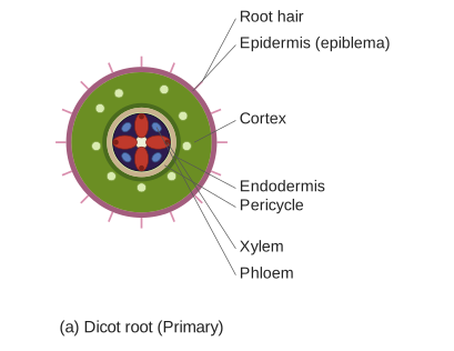
The primary structure of a dicot root, seen in transverse section, shows a characteristic radial arrangement of xylem and phloem, described here layer by layer since an image cannot be rendered as text.
A labelled transverse section of a young dicot root (e.g., of a bean or sunflower seedling) shows the following tissues arranged from the outermost to the innermost region: (1) Epiblema (also called the piliferous layer) — the outermost single layer of cells, some of which extend outward as unicellular root hairs for water/mineral absorption; it lacks a cuticle. (2) Cortex — several layers of thin-walled, loosely packed parenchyma cells with intercellular spaces, used mainly for storage; the innermost layer of the cortex is distinct. (3) Endodermis — a single layer of barrel-shaped cells forming the innermost boundary of the cortex; its radial and inner tangential walls bear a band of suberised, waterproof material called the Casparian strip, which forces water to pass through the endodermal cell cytoplasm (not between cells) on its way to the vascular tissue. (4) Pericycle — a single layer of thin-walled parenchymatous cells just inside the endodermis; it gives rise to lateral roots and is also involved in the origin of the vascular cambium during secondary growth. (5) Vascular bundles — arranged radially, meaning the xylem and phloem lie on different radii, alternating with each other, rather than forming a single conjoint bundle; the xylem is exarch (protoxylem lies tow …
Unlock everything free for 14 days
- Full step-by-step solutions
- Concept-first explanations
- Methods, shortcuts & mistakes
- PYQ mapping + timed mock tests
Full access for 14 days. No credit card required.