Q.Draw a well labelled diagram of T. S. Dicot root (Primary). OR Draw a well labelled diagram of T. S. of Monocot stem.
You're viewing a preview — the full solution, concept, methods & PYQ mapping are locked.
Start your 14-day free trial to unlock the full solution →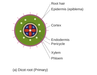
A dicot root's primary structure, viewed in transverse section, is arranged radially with epiblema outside, cortex and endodermis, pericycle, and a small number (2-4) of exarch xylem groups alternating with phloem, and little or no pith at the centre.
This question asks for a well-labelled diagram, which cannot be rendered as an image in this text answer — the internal anatomical arrangement it depicts is described below in full detail so the structure and labels are clear:
From outside to inside, a transverse section (T.S.) of a typical dicotyledonous root (primary structure) shows the following tissue regions, each of which would be a labelled part of the diagram:
- Epiblema (epidermis/piliferous layer): The outermost single layer of cells, without a cuticle or stomata, some cells of which bear unicellular root hairs that absorb water and minerals.
- Cortex: A wide zone of thin-walled, parenchymatous cells with intercellular spaces, located beneath the epiblema, mainly involved in storage.
- Endodermis: The innermost, single layer of the cortex, made of barrel-shaped cells without intercellular spaces, characterised by a band of suberised/lignified thickening on their radial and inner tangential walls, called the casparian strip.
- Pericycle: A single layer of cells just inside the endodermis, from which lateral roots and (later) the vascular cambium/cork cambium originate. …
Unlock everything free for 14 days
- Full step-by-step solutions
- Concept-first explanations
- Methods, shortcuts & mistakes
- PYQ mapping + timed mock tests
Full access for 14 days. No credit card required.