Chemistry · Ch 14 — Biomolecules
DNA double helix
DNA double helix
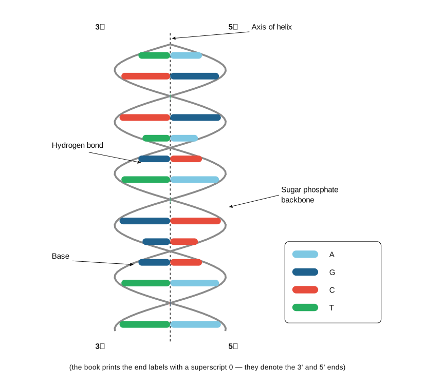
In 1953, the scientists James Watson and Francis Crick proposed a now-famous double-helix model for the overall three-dimensional structure of DNA, a model later confirmed directly by electron-microscope imaging. The salient structural features of the Watson-Crick DNA model are as follows. DNA consists of TWO separate polynucleotide strands, wound together around a common axis into a right-handed double helix. These two strands run in OPPOSITE chemical directions relative to one another -- they are described as antiparallel -- meaning that if one strand is read from its 5' end toward its 3' end, the second, partner strand running immediately alongside it is oriented the other way, from ITS 3' end toward ITS 5' end. Within this double-helical arrangement, the sugar-phosphate backbone of each strand lies on the OUTSIDE of the helix, while the nitrogenous bases of each strand point INWARD, into the interior of the helix, oriented roughly perpendicular to the helix's long central axis. The two strands are held firmly together, along the whole length of the double helix, by hydrogen bonding occurring specifically between paired bases on the two opposing strands -- this extensive, regular hydrogen bonding is what gives the DNA double helix its familiar 'ladder-like' internal structure, with the sugar-phosphate backbones as the ladder's two side-rails and the hydrogen-bonded base pairs as its regularly-spaced rungs. Crucially, this base-pairing is NOT random or arbitrary -- it follows a strict, universal complementary-pairing rule: adenine (A) always pairs specifically with thymine (T), forming exactly TWO hydrogen bonds between them, while guanine (G) always pairs specifically with cytosine (C), forming exactly THREE hydrogen bonds between them. Because of this strict A-T / G-C complementary pairing rule, the two strands of a DNA double helix are said to be complementary to one another: the complete base sequence of EITHER strand alone is enough to determine, unambiguously, the complete base …
Drawn by us to help you understand the concept clearly, and verified to make sure it's accurate. For exams, practice from your textbook's own diagram.
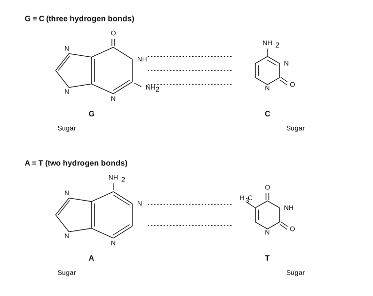
What this figure shows. A double-helical ladder diagram: two intertwined 'Sugar phosphate backbone' strands (drawn as the helix's outer rails, running in opposite 5'-to-3' directions, one strand's 5' end level with the other strand's 3' end) with an 'Axis of helix' marked down the centre, and repeated A-T and G-C base pairs shown as the ladder's rungs connecting the two backbones, each rung explicitly labelled 'Base' and 'Hydrogen bond'. An inset detail then draws out the actual hydrogen-bonding pattern atom-by-atom: cytosine (C) paired with guanine (G) via three hydrogen bonds (through the bases' respective N-H...O, N...H-N and O...H-N contacts), and thymine (T, bearing its characteristic -CH3 group) paired with adenine (A) via two hydrogen bonds (through N-H...N and O...H-N contacts) -- visually confirming the '2 bonds for A-T, 3 bonds for G-C' rule stated in the main text, and why …
Worked out. Worked problem: write the complementary strand for the DNA segment 5'-ACGTAC-3'. Solution: the complementary strand must run antiparallel (3' to 5' beneath the given 5'-to-3' original) and its bases must follow the A-T / C-G complementary pairing rule. Pairing each base of the original (A,C,G,T,A,C) gives, aligned directly beneath it, (T,G,C,A,T,G) -- so the complementary strand, written in its own aligned 3'-to-5' orientation, is 3'-TGCATG-5'. This worked method (align, then complement each base one-for-one, keeping the antiparallel orientation) is exactly the method used to answer Exercises 3.i …
Drawn by us to help you understand the concept clearly, and verified to make sure it's accurate. For exams, practice from your textbook's own diagram.
Why A pairs with T and G with C. The dashed lines are hydrogen bonds: G≡C forms three, A=T forms two. Only these combinations line up donor and acceptor atoms across the helix — the structural basis of complementarity that …