Q.Draw and label the areolar tissue.
You're viewing a preview — the full solution, concept, methods & PYQ mapping are locked.
Start your 14-day free trial to unlock the full solution →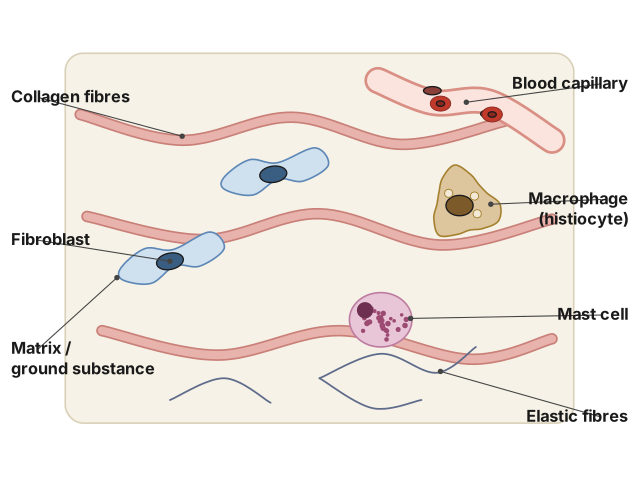
Areolar tissue is a loose connective tissue made of a jelly-like ground substance containing scattered fibres and cells such as fibroblasts, macrophages, and mast cells, forming a packing/binding tissue under the skin and around organs.
I cannot produce an actual hand-drawn diagram in this text format, so instead here is the structure of areolar tissue described part by part, exactly as it should be labelled in a diagram (this is the honest substitute for a drawing, per the platform's content rules). Areolar tissue is one of the most widespread loose connective tissues in the body, found beneath the skin (in the dermis) and around blood vessels, nerves, and organs, where it binds structures together while allowing some flexibility. In a labelled diagram of areolar tissue, the following components should be shown and named: (1) Matrix/ground substance — a semi-fluid, jelly-like intercellular substance in which the fibres and cells are embedded; (2) Collagen fibres — thick, wavy, glossy white fibres that provide tensile strength, scattered through the matrix; (3) Elastic fibres — thin, branching, yellowish fibres that provide elasticity, running among the collagen fibres; (4) Fibroblasts — the most abundant cells, flat and branched, responsible for secreting the fibres and ground substance; (5) Macrophages (histiocytes) — irregularly shaped phagocytic cells that engulf bacteria and debris, part of the tissue's defence role; (6) Mast cells — oval cells containing granules of heparin and histamine …
Unlock everything free for 14 days
- Full step-by-step solutions
- Concept-first explanations
- Methods, shortcuts & mistakes
- PYQ mapping + timed mock tests
Full access for 14 days. No credit card required.