Q.(a) Describe the structure of the human ovum with a neat labelled diagram. OR
You're viewing a preview — the full solution, concept, methods & PYQ mapping are locked.
Start your 14-day free trial to unlock the full solution →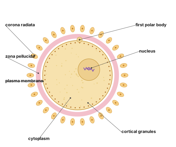
(a) The human ovum is a large, immotile secondary oocyte wrapped in the corona radiata and zona pellucida. (b) Populations are regulated toward their carrying capacity by density-dependent (biotic) and density-independent (abiotic) factors acting through birth, death, immigration and emigration.
(a) At ovulation, a mature Graafian follicle in the ovary ruptures and releases a large, spherical, non-motile secondary oocyte (commonly called the ovum), about 0.1 mm in diameter, arrested in metaphase of the second meiotic division (it completes meiosis II only if fertilised by a sperm). Structurally, immediately around the plasma membrane (oolemma) lies the cytoplasm (ooplasm), which is rich in stored nutrient/yolk granules and contains, just beneath the membrane, numerous small cortical granules that play a role in blocking polyspermy (fertilisation by more than one sperm) once the egg is fertilised. Surrounding the plasma membrane is a thick, transparent, non-cellular layer of glycoproteins called the zona pellucida, which sperm must penetrate to achieve fertilisation. Outside the zona pellucida is the corona radiata, a layer of follicle cells (granulosa cells) that remain attached to and radiate around the ovum after ovulation, having nourished and supported it during its growth in the follicle. Within the cytoplasm lies a large nucleus containing the chromosomes arrested at metaphase II, and the small first polar body (a byproduct of the first meiotic division) lies in the space between the oolemma and the zona pellucida. Unlike sperm, the human ovum lacks centrioles. …
Unlock everything free for 14 days
- Full step-by-step solutions
- Concept-first explanations
- Methods, shortcuts & mistakes
- PYQ mapping + timed mock tests
Full access for 14 days. No credit card required.