Q.Draw a T.S. of a young anther of an angiosperm. Label the different layers of the wall and write their functions.
You're viewing a preview — the full solution, concept, methods & PYQ mapping are locked.
Start your 14-day free trial to unlock the full solution →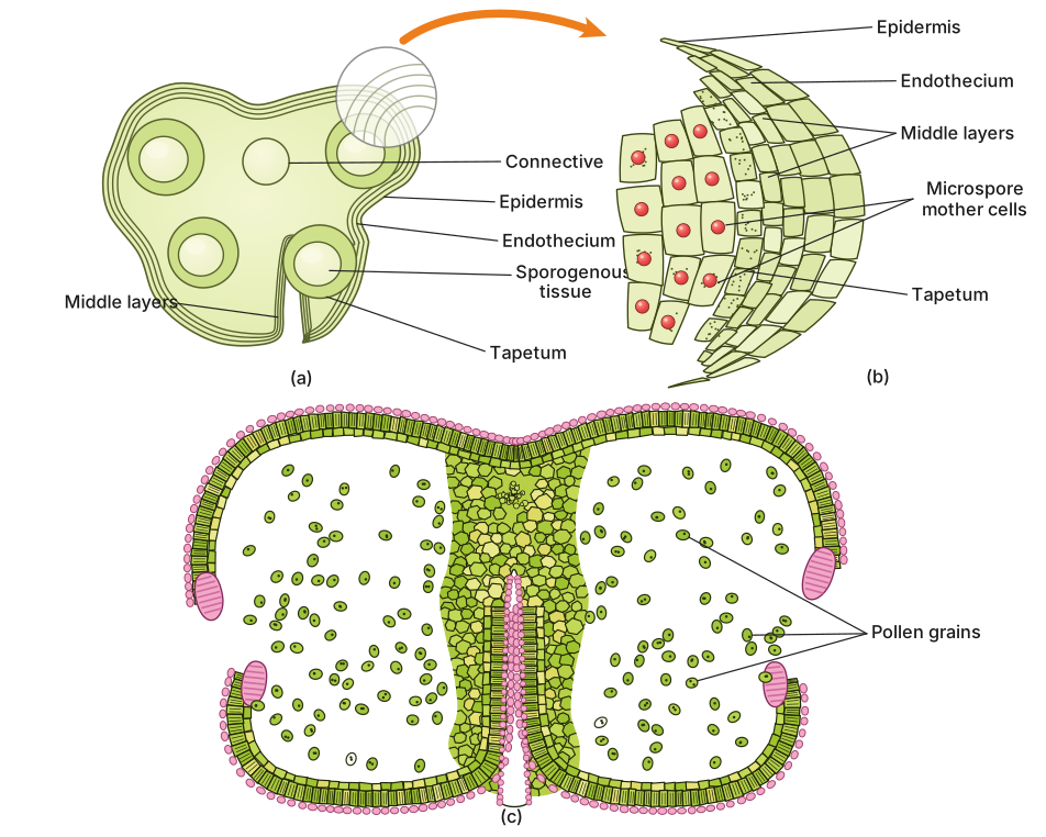
A young anther in transverse section shows four concentric wall layers — epidermis (protection), endothecium (dehiscence), middle layers (nutrition), and tapetum (pollen nourishment) — surrounding four pollen sacs (microsporangia) arranged in two lobes.
The anther is the male reproductive structure of a flower, and understanding its anatomy is essential to grasp how pollen grains develop and are eventually released. When you slice through a young anther transversely — that is, cut it across its width — you see a beautifully organized structure that reveals both the protective wall layers and the developing pollen sacs within.
Overall Architecture
A young anther appears bilobed in cross-section. Each lobe contains two pollen sacs (microsporangia), giving a total of four pollen sacs in a typical anther. These sacs are where the microspore mother cells will undergo meiosis to produce pollen grains. Surrounding these sacs is the anther wall, composed of four distinct layers, each with a specific role in protecting and nourishing the developing pollen.
The Four Wall Layers (from outside to inside)
1. Epidermis
The outermost protective layer, a single row of cells that forms a continuous skin around the anther. It guards the inner tissues against mechanical injury and water loss. In some species, the epidermal cells may develop into specialized structures, but primarily this layer serves as the first line of defense.
2. Endothecium
Lying just beneath the epidermis, this is a single layer of cells that develops fibrous thickenings on its inner tangential and radial walls as the anther matures. These thickenings are hygroscopic — they respond to changes in moisture — and play a crucial role in the dehiscence (splitting open) of the anther to release pollen. The endothecium essentially acts as the mechanical layer responsible for anther opening.
The fibrous bands in the endothecium are not present in very young anthers; they develop as the anther matures, which is why this layer is sometimes called the "fibrous layer."
3. Middle Layers
Between the endothecium and the innermost layer lie one to three layers of thin-walled cells. These middle layers are transient — they provide nourishment to the developing pollen sacs during early stages and are gradually crushed and absorbed as the anther matures and the pollen sacs expand.
4. Tapetum
The innermost layer, directly in contact with the developing microspores. The tapetum is the most metabolically active layer of the anther wall. Its cells are rich in nutrients and enzymes, and they nourish the developing pollen grains by secreting proteins, lipids, and the materials needed to form the pollen wall (including sporopollenin, the remarkably resistant substance that makes up the exine). The tapetum also contributes to the formation of the pollen coat. As pollen matures, the tapetal cells degenerate, their contents absorbed by the developing pollen. …
Unlock everything free for 14 days
- Full step-by-step solutions
- Concept-first explanations
- Methods, shortcuts & mistakes
- PYQ mapping + timed mock tests
Full access for 14 days. No credit card required.