Q.Draw a schematic transverse section of a mature anther of an angiosperm. Label its epidermis, middle layers, tapetum, endothecium, sporogenous tissue and the connective.
You're viewing a preview — the full solution, concept, methods & PYQ mapping are locked.
Start your 14-day free trial to unlock the full solution →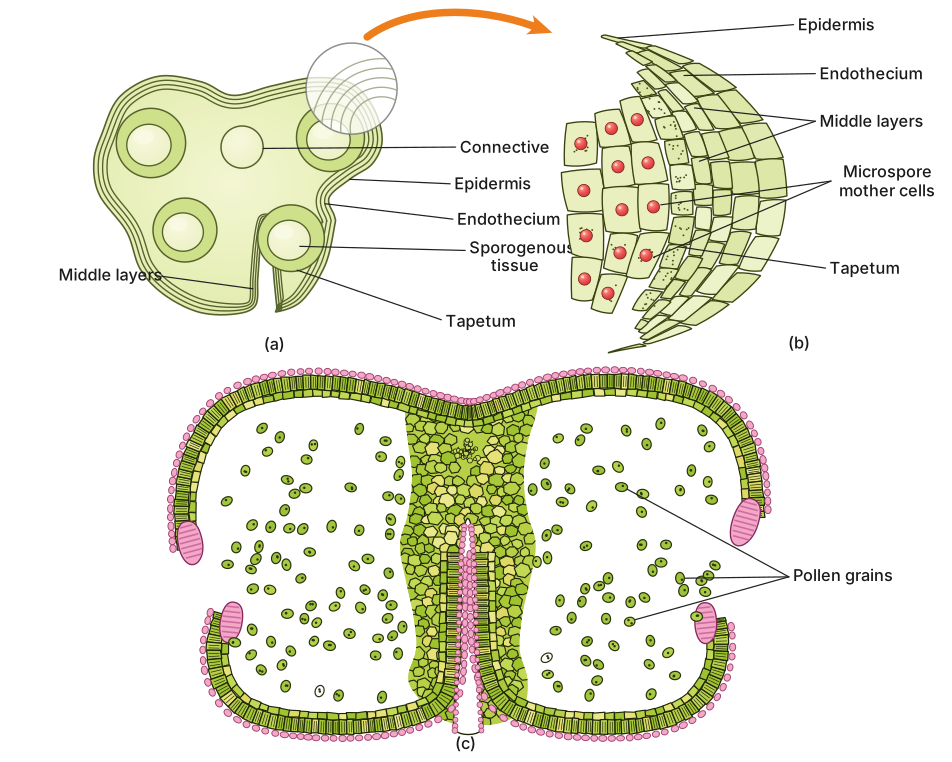
A mature anther in angiosperms is a four-lobed, bilobed structure whose transverse section reveals concentric wall layers — epidermis, endothecium, middle layers, and tapetum — surrounding the sporogenous tissue, all held together by a central connective.
To understand the anther's internal architecture, imagine cutting a mature anther crosswise — like slicing a tiny, four-chambered capsule. Each of the two lobes contains two pollen sacs (microsporangia), making four in total. The transverse section shows these four chambers arranged symmetrically around a central region.
The outermost layer is the epidermis — a single layer of protective cells that covers the entire anther. Just beneath it lies the endothecium, a layer of cells that develop fibrous thickenings at maturity. These thickenings are crucial: they help the anther dehiscence (split open) to release pollen. Below the endothecium, you'll find one to three layers of middle layers — thin-walled cells that often degenerate as the anther matures, providing nourishment temporarily.
The innermost wall layer is the tapetum, a specialised nutritive tissue that surrounds the sporogenous tissue. Tapetal cells are rich in nutrients and enzymes; they nourish the developing pollen grains and also contribute to the formation of the pollen wall. This layer is critical — without it, pollen development fails.
Inside the tapetum lies the sporogenous tissue, which consists of compactly arranged, densely cytoplasmic cells. These are the microspore mother cells (pollen mother cells) that undergo meiosis to produce haploid microspores, which then develop into pollen grains.
The sporogenous tissue is often described as "archesporial" in origin — it arises from a single layer of cells in the young anther that differentiates into the sporogenous mass.
Connecting the two anther lobes is the connective — a sterile, vascularised tissue that runs through the centre. It contains the anther's vascular bundle (xylem and phloem), which supplies water and nutrients. The connective also helps hold the four microsporangia together. …
Unlock everything free for 14 days
- Full step-by-step solutions
- Concept-first explanations
- Methods, shortcuts & mistakes
- PYQ mapping + timed mock tests
Full access for 14 days. No credit card required.