Biology · Ch 9 — Control and Co-ordination
The Ear: Structure, Hearing and Balance
The Ear: Structure, Hearing and Balance
Ear : The human ear -- the stato-acoustic organ -- performs two distinct jobs, hearing and maintaining body equilibrium, and is anatomically divided into external, middle and internal parts. The external ear consists of the pinna (an immovable, cartilage-supported structure in humans that collects sound and funnels it in), the auditory canal (which carries the sound to the eardrum, and whose skin bears fine hair and wax-secreting glands), and the tympanic membrane (eardrum) itself, a delicate structure of connective tissue covered by skin outside and mucous membrane inside that transmits the incoming vibrations to the middle ear.
The middle ear holds a chain of three ossicles -- malleus (hammer), incus (anvil) and stapes (stirrup, the smallest bone in the body) -- which amplify the eardrum's vibrations and pass them on to the cochlea; a short eustachian tube connects the middle ear to the pharynx and equalises air pressure on both sides of the eardrum. The internal ear consists of the labyrinth -- a bony labyrinth (filled with perilymph) enclosing a membranous labyrinth (filled with endolymph), the latter made of three parts: vestibule, semicircular ducts and cochlea.
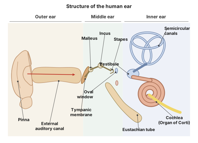
Drawn by us to help you understand the concept clearly, and verified to make sure it's accurate. For exams, practice from your textbook's own diagram.
What this figure shows. A full cross-section of the human ear showing the external auricle (pinna) and auditory canal leading to the eardrum; the air-filled middle-ear cavity containing the three ossicles malleus, incus and stapes seated in the oval window, with the auditory (eustachian) tube draining toward the throat; and the internal ear's bony and membranous labyrinth, comprising the coiled cochlea (with cochlear nerve), the vestibule (utricle and saccule) and the semicircular ducts (with vestibular nerve) opening via ampullae.
9.21: Structure of human ear. …
The cochlea, a coiled duct, holds three fluid-filled chambers separated by Reissner's membrane and the basilar membrane -- the upper scala vestibuli and lower scala tympani (both perilymph-filled) flanking the middle, endolymph-filled scala media. Sitting on the basilar membrane (the floor of the scala media) is the pea-sized organ of Corti, whose sensory epithelium of hair cells (bearing stiff microvilli called stereocilia) contacts an overlying gelatinous tectorial membrane, with supporting cells filling the gaps between hair-cell rows; this organ transduces sound vibrations into nerve impulses.
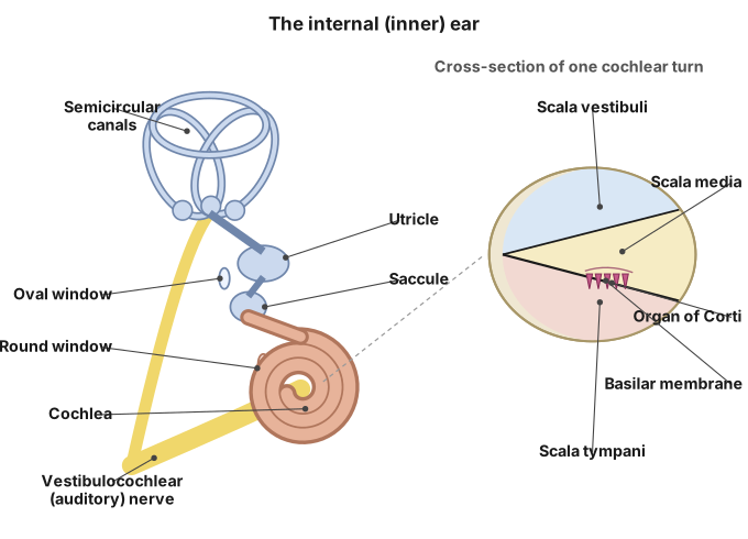
Drawn by us to help you understand the concept clearly, and verified to make sure it's accurate. For exams, practice from your textbook's own diagram.
What this figure shows. A close-up cross-section through one turn of the coiled cochlea showing the bony labyrinth surrounding the membranous cochlear duct, the vestibular canal (scala vestibuli) and tympanic canal (scala tympani) filled with perilymph on either side of the endolymph-filled cochlear duct, and the organ of Corti sitting on the basilar membrane, with its hair cells' stereocilia projecting up toward the overlying tectorial membrane and the auditory nerve fibres leaving at the base.
9.22: Human Internal Ear. …
Internal Ear and the mechanism of balance : Besides the cochlea, the inner ear's vestibular apparatus -- three semicircular ducts set at right angles to each other, plus the utriculo-saccular region with its otolith organ -- handles balance. Each semicircular canal's base has an ampulla containing a sensory crista, while the vestibule holds two sensory maculae (of the utricle and saccule); in both, hair-cell cilia project into a gelatinous otolithic membrane containing calcium-carbonate-and-protein granules (otoliths/otoconia). Cristae detect dynamic balance (movement), while the maculae of utricle and saccule detect static/linear balance (head position relative to gravity), and because the three canals lie in different planes, movement in any direction can be detected. …